12
HUMAN ANATOMY
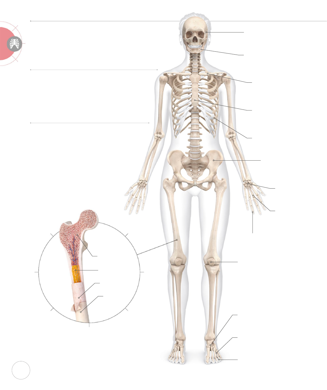
The 206 bones that make up your
skeleton are dynamic, living organs.
Together they form a framework for
your body that provides structure and
protection, and has the ability to move.
SYSTEM OVERVIEW
Your bones are made of collagen and they
store calcium, a mineral that makes them
strong and is vital for bodily functions. They
also contain bone marrow where blood
cells are produced. Bones form joints,
which are supported by cartilage and
structures such as ligaments. Yoga can
support your bone and joint health.
SKELETAL
SYSTEM
Bone structure
Bone has a smooth outer connective tissue
shell called periosteum. Inside this is a
strong, dense layer known as compact bone.
Deeper still is spongy bone with
honeycomblike spaces; this is strong yet light.
Mandible
Lower jawbone that
forms the only movable
joint in your skull
Clavicle
Also called the collar-
bone, it connects your
scapulae and sternum
Carpals
Eight small bones
form each wrist
Metacarpals
Five long bones run
through each palm
Phalanges
Each hand has
14 bones forming
your fingers
Sternum
Also called the
breastbone, it
connects your ribs
Ribs
The 12 pairs of bones
that form your ribcage
Pelvis
Two hip bones
connected by
your sacrum
Tarsals
The seven small bones
that form your ankle
Patella
Also called the kneecap,
it is attached to your
quadriceps tendon
Metatarsals
Five long bones that
run through your foot
Phalanges
The 14 bones in each
foot that form your toes
Skull
These fused plates of
bone protect your brain
Periosteum
Compact bone
Bone marrow
Spongy bone
US_012-013_Skeletal_01.indd 12 02/11/2018 14:00

13
Cartilage
Hyaline articular cartilage lines bones at most
joints and is smoother than glass—it even
looks like stained glass under a microscope.
However, when this cartilage wears down, it
can become coarse like sandpaper, causing
a condition called osteoarthritis (see p.17).
Ligaments
Bones are connected by dense bers called
ligaments. Both ligaments and tendons (see
p.19) have very little elasticity, meaning, if you
overstretch them in an asana, they often don’t
go back to their resting length and lose stability.
Vertebral column
A series of bony disks
that form your spine
Scapula
The shoulder blade
connects torso and arm
Humerus
This bone connects your
scapula and forearm
Sacrum
This bone is the
keystone of your pelvis
Ulna
Forearm bone
that runs to your
little finger
Radius
Forearm bone
that runs to
your thumb
Femur
Your thighbone
is the longest bone
in your body
Tibia
You can feel the edge
of your shinbone
under your skin
Fibula
Thin bone that sits on
the outside of your leg
Calcaneus
Your heel bone attaches
to your Achilles tendon
Ligament
attaches bone
to bone
Chondrocyte
(cartilage cell)
US_012-013_Skeletal_01.indd 13 02/11/2018 14:00

14
SPINE
Your vertebrae sit on top of each other to create natural curves.
This is called a “neutral spine.” It alternates between curving
inward (lordosis) and outward (kyphosis) to absorb shock like
a coiled spring. Your vertebrae are like wedges stacked to form
these curves in order to bear your body weight most eciently.
Cervical
curvature
There is
a natural
lordosis in
the neck
Thoracic
curvature
There is a natural
kyphosis in the
mid-back
Lumbar
curvature
There is
a natural
lordosis in the
lower back
NEUTRAL SPINE
These natural curves
create the strongest, most
stable alignment of the
spine. In this ideal, the
spine is also not twisted
or leaning to either side.
KYPHOSIS
Hyperkyphosis of the
thoracic spine is often
simply called a kyphosis
or hunchback. This
exaggerated curvature is
common in osteoporosis.
LORDOSIS
Hyperlordosis of the
lumbar spine is sometimes
just called a lordosis
or swayback. This
exaggerated curvature is
natural during pregnancy.
Neutral spine
Many asanas incorporate a neutral spine,
such as seated meditation poses. Poor
posture and other considerations can
lead to a multitude of spinal structural
deviations, including common conditions
like hyperlordosis and hyperkyphosis.
Yoga works your spine in unique ways
and enhances body awareness to
improve your overall posture.
THORACIC VERTEBRA
LUMBAR VERTEBRA
CERVICAL VERTEBRA
Gentle, even curves
Curvature in upper
spinal column
Curvature in lower
spinal column
Space for
vertebral
artery
Hole for
spinal cord
Body of
vertebra
Articulates
with rib
Enlarged body to
support weight
Articular
process
US_014-015_Skeletal_02.indd 14 02/11/2018 14:00
15
HUMAN ANATOMY
Skeletal System
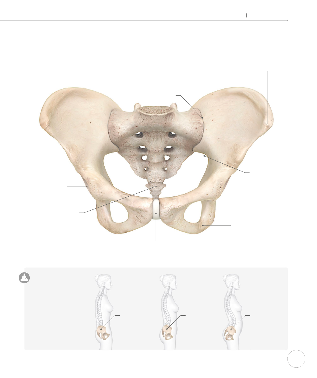
PELVIS
Your pelvis includes two hip (coxal) bones connected by your
sacrum. The sacrum, which means “sacred” in Latin, is the
triangular bone with the tailbone at the lower, or inferior,
end; it acts like the keystone to an arched bridge, forming
a structurally sound base for your spine.
Acetabulum
The socket of your hip
joint which articulates
with your femur
Coccyx
These fused bones are
known as the tailbone
Pubic symphysis
This joint is made of
fibrocartilage like your
intervertebral disks
Sacroiliac joint
Commonly called
the SI joint, this is
slightly movable
Ischial tuberosity
Your “sitting bones”
are at the base of
your pelvis
Greater sciatic notch
This creates a space for
the sciatic nerve to pass
Neutral pelvis
A neutral pelvis facilitates a
neutral spine and vice versa.
Imagine your pelvic bowl
lled with water. Finding a
neutral spine and pelvis
means that the water
wouldn’t spill backward,
forward, or to the side—
such as when one of your
hip points is lifted or
your pelvis is rotated.
Anterior superior
iliac spine
Your “hip points” can be
felt under your skin
FEMALE PELVIS
POSTERIOR TILT
Pelvic bowl
tilts backward,
flattening
lumbar curve
NEUTRAL
Pelvic bowl
is balanced,
with neutral
lumbar curve
ANTERIOR TILT
Pelvic bowl
tilts forward,
exaggerating
lumbar curve
US_014-015_Skeletal_02.indd 15 02/11/2018 14:00

16
JOINTS
Joints are where bones unite and articulate to allow movement.
There are three joint types: fibrous, cartilaginous, and synovial.
Fibrous joints are immobile, such as the sutures in your skull.
Cartilaginous joints are slightly mobile, like your pubic symphysis.
Synovial joints are most mobile and are very important for asanas.
Inside a joint
Synovial uid lubricates and cushions. It
is a “non-Newtonian uid,” which means
it gets more viscous or thicker in response
to pressure, similar to solutions of cornstarch
in water. With a sedentary lifestyle, synovial
uid may become thin and less eective.
However, impact from the practice of yoga
asanas causes synovial uid to thicken,
reducing pain and better protecting joint
structures such as cartilage.
JOINT ACTIONS
Synovial joints of your body can move in many
directions. Hinge joints in your elbow and knee
mainly perform flexion and extension, like the
hinge of a door. Larger ball and socket joints like in
your shoulder and hip can also perform abduction,
adduction, rotation, and circumduction, which is
a combination of all of the above movements.
SYNOVIAL JOINT
Synovial joints allow movement while
protecting bone ends from touching
each other, which would cause damage.
They are the most common type of
joint in the body. ELBOW JOINT
TRIANGLE
Hip external
rotation
Hip internal
rotation and
abduction
Elbow extension
Shoulder
abduction
Spinal axial
rotation
Ligaments
connect bone
to bone
Bones come
together to
create a joint
Spongy bone
Healthy synovial
fluid is thick to
cushion joint
Articular cartilage
reduces friction
Joint capsule
holds joint
together
Knee extension
Shoulder
external
rotation
TYPES OF MOVEMENT
Flexion
Angle at joint generally gets smaller
Extension
Angle at joint generally gets larger
Abduction
A limb moves away from the body
Adduction
A limb moves closer toward
the body
External rotation
A limb rotates outward
Internal rotation
A limb rotates inward
Axial rotation
The spine twists on its axis
Plantar exion
Pointing the feet
Dorsiexion
Flexing the feet
US_016-017_Skeletal_03.indd 16 02/11/2018 14:01
..................Content has been hidden....................
You can't read the all page of ebook, please click here login for view all page.