
129Zero to Genetic Engineering Hero - Chapter 5 - Extracting your engineered proteins
A. Using your DNA Playground or a tube rack, set in the Final Pigment Tube and remove the lid.
B. Remove the syringe plunger from the syringe and lay it on a clean surface.
C.
Open up the syringe lter packaging but DO NOT fully remove the lter from its packaging. Do this by either:
a) taking the paper cover off if the lter is in a plastic/paper package, or, b) by opening the sealed plastic bag
it is in. You do not remove the lter because you want to make sure that you do not contaminate it before use.
D. Holding the lter via the plastic container/bag, screw on the syringe to the lter, so it is rmly connected.
You can lay this on the table, but be sure not to touch the sterile output end of the lter.
E. Extra Protection: Use the Burst Bag in the Plate Extract-it Kit to enclose the syringe and lter. The Burst
bag is not a foolproof solution, but in the rare event that a burst event happens, the bag will act as the rst line
of blockage for a spraying sample.
F. After prepping your Burst Bag, and placing your syringe/lter inside it, gently pour or pipet your Lysis
sample into the open syringe. Do this gently to avoid causing the pellet of cell debris from falling into the syringe.
If this happens, the pellet will clog up the syringe. If you see clumps of cell debris pour into the syringe, pour
all the contents back into the tube and repeat the centrifugation step.
G. With the sample in the syringe, hold it so that the sterile end of the lter points into the Final Pigment/
Product Tube. Replace the syringe plunger into the syringe and GENTLY but FIRMLY press down. If you have
effectively microcentrifuged your sample, the plunger will slowly push in until all the solution passes through.
Cell debris or bacteria in the sample will be trapped in the lter. Small molecules like your proteins will pass
through. In the event that most but not all liquid passes through before cloggging the lter, this tells you that
next time, you should incubate in the lysis tube longer or centrifuge a bit longer.
H. Close and tighten the lid on your Final Product Tube. Congratulations! You have now sterilized the proteins
you microfactured using your genetic engineering skills! You can store your nal pigment in the refrigerator,
or at room temperature. Many color pigments will keep their color for more than a year if kept out of the sun.
Step 11. Using your proteins
Figure 5-10. Examples of uorescent pigments extracted from colored proteins engineered in E. coli with Amino Labs Engineer-it Kit
and Extract-it Kit: i) Fluorescent proteins under UV light; ii) Etched plastic dyed with uorescent Yellow, Cyan, and Magenta proteins
by J. Pahara, 2017; iii) Frog painting made with Fluorescent Yellow and Cyan extracted proteins by John from Toronto, Canada, 2016
Book _genetic engineering hero-AUG2021.indb 129Book _genetic engineering hero-AUG2021.indb 129 8/18/21 12:03 PM8/18/21 12:03 PM

130 Zero to Genetic Engineering Hero - Chapter 5 - Extracting your engineered proteins
on completing your fourth experiment!
This cell extract with a high concentration of the colored protein can be used in different ways (Figure 5-10):
• If it is a uorescent protein, place it next to a black light to see it glow
•
Express yourself artistically! Try using it as ink or watercolor. You can use different types of paper and
drawing instruments like paintbrushes. If you have a uorescing protein and plan to use a black light to
illuminate your artwork, note that a black light can cause some light papers to glow blue, which may affect
your artwork; place a black light next to the paper to test before.
• Wear your sample proudly, or even gift it: Find a small container that can hold liquid. Seal the opening and
attach the container to a necklace, pin or other wearable item!
• Try dyeing fabrics or other materials with it! You can even layer it onto etched or porous plastic. See if you
can dye that surface!
Book _genetic engineering hero-AUG2021.indb 130Book _genetic engineering hero-AUG2021.indb 130 8/18/21 12:03 PM8/18/21 12:03 PM
131Zero to Genetic Engineering Hero - Chapter 5 - Extracting your engineered proteins
Fundamentals: How cells translate proteins from RNA
Step two of the Three Steps
to Microfacturing: Translating
proteins from RNA
Let’s pick up from where we left off in Chapter 4. In
your hands-on experiment, when you inserted DNA
plasmids into cells, RNA transcripts of genes were
transcribed from DNA templates by RNA polymerases.
Depending on the sigma factor and how strongly it
bound to the promoter, there could be tens, hundreds,
or thousands of RNA transcripts from your gene of
interest bumping around the K12 E. coli cell. Each
transcript becomes a key part of the second step of
Microfacturing - translation.
Just like how RNA polymerase “reads” DNA, binds
ribonucleotides and strings them together to form a
strand of RNA, translation involves a different cellular
machine called a ribosome that binds to and “reads”
RNA. While reading the RNA, it binds up amino acids
and strings them together to form proteins. If you’ve
forgotten what amino acids are, check back to Chap-
ter 3. Also, just like we saw in Chapter 4 how DNA has
coding and non-coding regions, RNA also has coding
and non-coding regions (Table 5-1). These regions
determine when and how much protein is translated
an
d where to start translating from.
The equivalent of a promoter in DNA is the ribo-
somal binding site (RBS) in RNA. In other words, in a
DNA molecule, the promoter helps the transcription
machinery know where to start transcription, and
in an RNA molecule, the RBS helps the translation
machinery to know where to start translation.
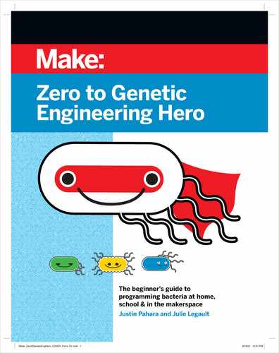
In Figure 5-11, notice that DNA contains all the infor-
mation for transcription and translation: Promoter,
RBS, and the coding sequence. As mentioned in Chap-
ter 1, DNA is the master blueprint of the cell. During
transcription, embedded in the DNA coding region is
an RBS - the RBS does not have any function during
transcription. Only once the RNA transcript is
created,
will the RBS, which is now at the 5’ phosphate end
of the RNA transcript, will become relevant and be
Table 5-1. Non-coding regions that function in DNA and RNA
Nucleic Acid Non-coding region Position Function
DNA Promoter
The promoter is upstream of the RNA
coding region and determines the
DNA strand that the RNA polymerase
transcribes.
Binds to sigma factors which then can
bind to and orient RNA polymerase
to commence the creation of RNA
transcripts through the process of tran-
scription
RNA
Ribosomal binding
site (RBS)
In DNA, the RBS is just downstream
of the promoter but is still upstream
of the protein coding region. In RNA
the RBS is at the 5’P end of the tran-
script.
Binds to initiation factors which then can
bind to the ribosome and can commence
the creation of proteins through the
process of translation
Figure 5-11. During the rst two steps of the Three Steps to Mi-
crofacturing, DNA and RNA each contain coding and non-cod-
ing regions. Upon the creation of the protein chain during
transcription, it folds into a 3D structure called a protein. It
is the 3D structure of the protein that determines its function.
Coding Sequence to Produce Protein
Coding Sequence to Produce RNA
Promoter RBS
RBS
DNA
RNA
transcript
Protein
(amino acid chain)
Functional 3D Protein
Translation
Protein Folding
Transcription
Book _genetic engineering hero-AUG2021.indb 131Book _genetic engineering hero-AUG2021.indb 131 8/18/21 12:03 PM8/18/21 12:03 PM

132 Zero to Genetic Engineering Hero - Chapter 5 - Extracting your engineered proteins
key in causing translation to start. The region down-
stream of the RBS is the RNA coding region, which
codes for the protein to be created by the translation
machinery.
During translation, there is the creation of the amino
acid chain (protein). The folding of that chain into a
three-dimensional shape results in the protein struc-
ture that has a function.
Starting Translation
How does the cell machinery know when and how
to start translating an RNA transcript? Transla-
tion involves similar principles as in transcription:
Proteins are bumping around the cell and can bind
to the ribosomal binding site in RNA, which can then
also bind to a ribosome - these protein are called initi-
ation factors.
Proteins already made by the cell called initiation
factor 1 (IF1), initiation factor 2 (IF2), and initiation
factor 3 (IF3) bump around in the cell. While these are
three different proteins, they all work together to start
the process of translation. In accordance with the
Four B’s, the initiation factors bump around until they
interact with the ribosome. The ribosome’s purpose is
to read the RNA transcript sequence and translate the
RNA sequence into an amino acid chain. An amino
acid chain is commonly referred to as a protein. Just
like RNA polymerase, the ribosome will use a cipher
to do this, which we will explore in the next section.
The initiation factors also help to start translation
by binding to the RNA transcript. If the ribosomal
binding site (RBS) has the right shape and charge to
bind to one or more initiation factors, the initiation
factor(s) will bind.
For the translation process to be successful, when the
ribosome binds to the RNA via the initiation factors,
it must also lock into the RNA so that it does not “fall
off”. This is similar to how, during transcription, the
RNA polymerase created a short piece of RNA called
the initiation sequence. However, the ribosome is
slightly different thanks to a special “built-in” feature
that pre-equips it with the locking mechanism. As you
can see in Figure 5-12, the ribosome is made up of
both protein and RNA intertwined with one another.
That’s right, the ribosome is actually a hybrid of both
protein and RNA! It is the ribosomal RNA that allows
it to lock into the RNA strand through complementary
ribonucleotide interactions (A-U, G-C) (Figure 5-12,
Figure 5-13).
Figure 5-12. A crystal structure of a ribosome. Ribosomes
bind to and read RNA and translate their sequence into a se-
quence of amino acids - also called a protein. The ribosome
itself is a mixture of both protein and RNA that function in
harmony. Purple: “16s RNA” strand; Red: other RNA strands;
Blue: Protein. Crystal structure data from A. Korostelev, S.
Trakhanov, M. Laurberg and H.F. Noller (2006) Crystal Struc-
ture of a 70S Ribosome-tRNA Complex Reveals Functional In-
teractions and Rearrangements. Cell 126:1065-1077.
Figure 5-13. The ribosome is made up of both protein (blue)
and RNA (yellow). It is the ribosomal RNA (rRNA, yellow) that
allows the ribosome to lock into the RNA transcript (orange)
by complementing the ribosomal binding site (RBS) at the 5’P
end of the RNA transcript (orange).
rRNA
Ribosome (Protein)
RBS
RNA transcript
Book _genetic engineering hero-AUG2021.indb 132Book _genetic engineering hero-AUG2021.indb 132 8/18/21 12:03 PM8/18/21 12:03 PM

133Zero to Genetic Engineering Hero - Chapter 5 - Extracting your engineered proteins
This ribosomal RNA (rRNA) is known as 16s rRNA
and is intertwined within the protein structure of the
ribosome. 16s rRNA is normal RNA, except it doesn’t
get translated into a protein, and simply stays as RNA.
The rRNA folds upon itself in a way that allows it to
also merge with the ribosome protein (Figure 5-12).
The rRNA is an essential part of the ribosome because
it is what enables the protein structure to lock into
the RNA transcript thanks to complementary binding
of ribonucleotides. While the initiation factors help
the ribosome bind to the RNA initially, it is the 16s
rRNA that gets the ribosome in position and readies
it for translation. Just like two complementary DNA
strands can come together, the rRNA complements
a short sequence of the ribosomal binding site of the
RNA transcript (Figure 5-13).
During Translation: The RNA to
protein cipher
The initiation factors do the Four B’s and bind to the
ribosome, which further bumps around and becomes
bound to the RNA transcript at the RBS. The rRNA in
the ribosome complements and bonds to the RBS of
the RNA transcript. Now that the ribosome is locked
into the RNA at the RBS using the rRNA, it can start the
process of translation. Translation involves “reading”
the RNA transcript while simultaneously creating an
amino acid string.
Let’s briey revisit how RNA polymerase works during
transcription, as it has some simimlarities to how the
ribosome works in translation. The RNA polymerase
uses a cipher to “read” DNA and transcribe RNA
(Table 4-2). The RNA polymerase cipher is based on
the complementarity of the DNA and the ribonucle-
otides (A’s bind to U’s, C’s bind to G’s, T’s bind to A’s).
While the RNA polymerase “reads” the DNA, millions
of ribonucleotide molecules (A’s, U’s, G’s, C’s) bump
around and, when the “right” ribonucleotide “t in”
to the RNA polymerase and complements the DNA
nucleotide being read by the polymerase, the RNA
polymerase permanently attaches it to the growing
chain (Figure 4-29).
Translation also has a cipher that relies on complmen-
tarity and the Four B’s, but is slightly more complicated
than the one for transcription. Let’s explore it now,
along with the machinery that the ribosome uses to
“read” RNA and create a chain of amino acids. Unlike
in transcription, where the RNA polymerase simply
“adds on” ribonucleotides that complement the nucle-
otides in the DNA template strand, amino acids cannot
directly complement the RNA transcript. Therefore,
the ribosome needs a “go-between” to bridge the gap
between the RNA transcript and the amino acid. These
“go-betweens” are another kind of hybrid molecule
called transfer RNA (tRNA) (Figure 5-14). tRNA is a
hybrid molecule made up mostly of RNA and one
amino acid: the one end of the tRNA is able to inter-
act with and complement the RNA transcript through
what is called an anticodon, and on the other end is an
amino acid that the ribosome can add onto the grow-
ing amino acid chain. Coincidentally, the tRNA also
sort of has a hand-written cursive capital “T” shape
(Figure 5-14).
Quite a lot happens in translation in order for the
RNA to be translated into a protein. Let’s pause and
summarize all of the different players involved in
translation:
•
RNA transcript/messenger RNA: The RNA
polymerase, a protein enzyme, transcribed the
information from DNA to make the RNA tran-
script during transcription. Another name for the
RNA transcript is messenger RNA (mRNA). The
RNA transcript has a non-coding region called a
ribosomal binding site (RBS), as well as a coding
region which is what will ultimately be translated
into a protein.
•
Initiation factors: Initiation factors bind to the
ribosome and RBS of the RNA transcript. They are
analogous to the sigma factors in transcription.
Figure 5-14. A transfer RNA (tRNA) molecule is a string of
non-coding RNA that folds into a “T” shape and has an amino
acid at one end while the other end binds to the RNA tran-
script. It folds into this shape simply because of complemen-
tary regions of RNA nucleotides. See the hairpin structure of
an RNA terminator in Chapter 4.
Amino Acid
Anticodon
Book _genetic engineering hero-AUG2021.indb 133Book _genetic engineering hero-AUG2021.indb 133 8/18/21 12:03 PM8/18/21 12:03 PM
..................Content has been hidden....................
You can't read the all page of ebook, please click here login for view all page.