7
Early Prediction of Epileptic Seizure Using Deep Learning Algorithm
T. Jagadesh1*, A. Reethika1, B. Jaishankar1 and M.S. Kanivarshini2
1Department of Electronics and Communication Engineering , KPR Institute of Engineering and Technology, Coimbatore, Tamil Nadu, India
2HCL Technologies, Chennai, Tamil Nadu, India
Abstract
Epilepsy is one of the most common neurological disorders in the world. Early expectation remains based on approach of seizures affects existence with epileptic patients. A novel patient-explicit seizure is defined in this article, expectation procedure dependent on profound learning and functional to extended haul scalp electroencephalograms (EEG) chronicles is proposed. The main objective is to recognize the preictal mind state besides separate it from predominant at the state of interictal at the correct time as could be expected and make it reasonable for continuous. In this highlights, extraction and characterization measures were consolidated into a solitary computerized framework. Crude EEG signal with no pre-processing is well-thought-out as contribution to the framework, further lessens the calculations. Four profound learning models have been proposed to extricate the most judicial highlights that upgrade the order exactness and expectation time. Our new approach exploits the convolutional neuronic organization in removing the huge spatial highlights from various scalp positions and the repetitive neural organization in expecting the frequency of seizures sooner than the current techniques. A semi-directed methodology dependent on exchange learning strategy is acquainted with enhancement issue. A channel choice calculation is proposed to choose the most pertinent EEG, which makes the projected framework great possibility for continuous use. A powerful test technique is used to guarantee vigor.
Keywords: Epileptic seizure, deep learning, EEG signal
7.1 Introduction
Seizures are a symptom of a primary brain disorder that might short-term or long-term. These arise by chemical changes in nerve cells causing an aberrant burst of electrical commotion in the brain. In most cases, excitant and repressive brain cells are in equilibrium. Former encourages action, whereas latter discourages. When this equilibrium is disturbed and there is either too much or too little activity, seizures (as shown in Figure 7.1) can develop.
Reasons for Seizure Disorders
Seizures consist of many factors:
- Hypoxic-ischemic encephalopathy (HIE)
- Birth injury/trauma
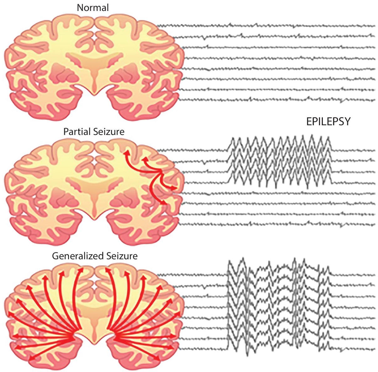
Figure 7.1 Seizure identification.
- Infection
- Genetic factors
- Metabolic/chemical imbalances
- Medication side effects
- Brain tumors
Twitching, spasms, and loss of consciousness are all symptoms of seizures. Changes in feelings and behavior are also possible. It’s crucial to understand that seizures can be mild and don’t always include the spectacular convulsions that most people associate with them. If a parent or guardian suspects their kid is experiencing a seizure, they should seek medical help immediately.
Seizure disorder diagnostic, such as:
- Electroencephalogram (EEG)
- Bloodwork
- Computed tomography scan (CT or CAT scan)
- Magnetic resonance imaging (MRI)
- Lumbar puncture (spinal tap)
Epilepsy is formed by gathering of nervous problems portrayed by intermittent epileptic seizures [10, 11], scenes can be changed from ephemeral and almost imperceptible stages of extensive stretches to enthusiastic trembling because of unusual electrical movement in the brain [1]. These scenes can bring about actual wounds, either straight-forwardly like broken bones or through causing accidents [1]. In epilepsy, seizures tend to repeat and have no prompt basic reason [10]. Isolated seizures triggered by a specific cause, such as harming, are not thought to be epilepsy [12]. People affected by epilepsy can treated in various method and feel vary degrees of social shame as a result of their diagnosis [1]. Extreme and strange synaptic action in the cortex of the brain is the secret weapon of epileptic seizures [12]. The cause of epilepsy remains unexplained much of the time [1] a few results (Figure 7.2 shows all the types of results) of brain damage, cerebrum cancers, cerebrum defects, or birth deserts in an epileptogenic period [1–3]. Known hereditary transformations are straightforwardly connected to a little extent of cases [4, 13]. The analysis includes precluding different conditions that may cause comparable indications, for example, swooning, and deciding whether another reason for seizures is available, like liquor taking away or electrolyte problems [4]. This might be a part of the way done by the imaging mind and blood tests.
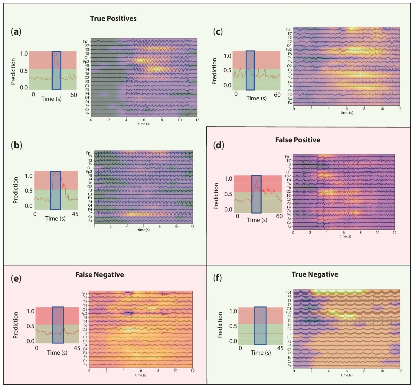
Figure 7.2 Seizure detection simulation results.
Epilepsy that happens because of different issues might curable [1]. Controllable with prescription in the range of 70% cases [7]; cheap enemy of seizure drugs are regularly available [1]. In those whose seizures don’t react to medicine, medical procedure, dietary changes may be considered [5, 6]. It is not considered in all instances of deep rooted epilepsy, numerous individuals to point therapy [1].
The most widely recognized sort (60%) of seizures is convulsive which include compulsory muscle contractions [23]; 33% start as summed up seizures from the beginning, influencing the two sides of the equator of the mind and impeding consciousness [23]; 66% start as central seizures (which influence one half of the globe of the cerebrum) advance to summed up [23]. The excess 40% of seizures are non-convulsive. An illustration of sort is the nonattendance seizure, which presents as a diminished degree of cognizance and ordinarily keeps going around 10 seconds [24].
Central seizures are frequently gone before by specific encounters, known as auras. Seizures incorporate tactile (visual, hearing, or smell), clairvoyant, autonomic and engine wonders relying upon what portion of the cerebrum is involved [2]. Muscle jerks may begin in a particular muscle gathering and spread to encompassing bunches, it is known as a Jacksonian march. Automatisms may happen, which are non-deliberately created exercises, for the most part basic dull developments like resembling the lips or more mind boggling exercises, for example, endeavors to get something.
There are six primary kinds of summed up seizures: tonic-clonic, tonic, myoclonic, nonappearance, and unaccented seizures. They all include loss of cognizance and ordinarily occur abruptly.
Tonic-clonic happens in constriction of appendages tracked by their expansion alongside angling of the back which keeps going 10–30 seconds (tonic stage). An exclamation cry might hear because of the constriction of chest muscles, trailed by a shuddering of the appendages as one (clonic stage). Tonic seizures yield consistent compressions of muscles. An individual regularly becomes blue as conscious is halted. In clonic seizures, there is shaking of appendages as one. After the shuddering, halted might require 10–30 minutes for an individual to get back to the business as usual; this is known as “postictal state”. Damage inside or bladder control might happen during seizure. Society encountering a seizure stay quiet, either the tip or on the sides; tonic-clonic seizure, chomps sides are added common. Tongue nibbles additionally moderately basic psychogenic non-epileptic seizures.
Myoclonic seizures include fleeting fits of muscles in a couple of regions or all over. Occasionally, it causes the individual to fall, which may get injury. Absence of seizures can be unpretentious with a slight turn of the head or eye squinting with weakened mindfulness [2]; regularly, individual doesn’t drop over and gets back to its ordinary at it ends [2]. Atonic seizures includes the deficiency of muscle movement for more than 1 second, normally happening at the two sides of body. Scarcer seizure can cause compulsory abnormal snickering crying, or further perplexing encounters, for example, déjà vu.
About 6% of epilepsy have seizures were regularly set off by explicit occasions known as reflex seizures. With reflex epilepsy are just set off by explicit stimuli. Communal triggers incorporate blazing illuminations and unexpected sounds. In particular sorts of epilepsy frequently during sleep, and in different kinds it happens practically just while sleeping.
Assuming seizures emerge from a particular space of the cerebrum, the underlying indications of the seizure regularly mirror the elements of that space. The correct portion of the mind control the left half of our body, and the left 50% of the cerebrum control the correct side of our body. For instance, if a seizure begins from the correct side of the cerebrum in the space that controls development in the thumb, at that point the seizure may start with jolting of the left thumb or hand.
Seizures differ such that a lot of epilepsy experts habitually rename seizure types. Commonly, seizures have a place in one of two fundamental classifications: essential summed up and incomplete seizures. The contrast can be analyzed between the sorts of the way start. Essential summed up seizures start with a far-reaching electrical release that includes the two sides of the cerebrum immediately. Halfway seizures start with an electrical release in one restricted space of the cerebrum.
Epilepsy in which the seizures start from the two sides of the mind simultaneously is called essential summed up epilepsy. Innate elements are significant in fractional summed up epilepsy, which is bound to include hereditary variables than incomplete epilepsy – a condition where the seizures emerge from a restricted space of the mind.
Some fractional seizures are identified with head injury, mind contamination, stroke or tumor be that as it may, much of the time, the reason is obscure. One inquiry that is utilized to additionally order incomplete seizures is whether awareness (the capacity to react and recall) is hindered or protected. The distinction may appear glaringly evident, yet there are numerous levels of cognizance impedance or conservation.
Epilepsy might be treated with antiepileptic drugs (AEDs), diet treatment, and medical procedure. Prescriptions are the underlying treatment decision for practically all patients with different seizures. A few patients who just have a solitary seizure and whose tests don’t show a high probability of seizure repeat may not need drugs. The meds treat the manifestations of epilepsy (the seizures), instead of relieving the fundamental condition. They are exceptionally successful and totally control seizures in the dominant part (roughly 70%) of patients. The medications keep seizures from beginning by lessening the inclination of synapses to convey unreasonable and confounded electrical messages.
Profound learning has been generally utilized for computerized EEG preparing in various settings, for example, mind PC interfaces [7–10], programmed rest scoring [11–14] and epileptic seizure prediction [15–17], and detection [18–22], due to its ability to take in rich portrayals from crude data [23] and its fruitful presentation on visual acknowledgment undertakings, particularly on common images [24]. Specifically, for the programmed seizure identification issue, late methodologies are summed up in Table 7.1. In this table, the initial two sections allude to the technique, the third to the data set utilized for the assessment, and the last ones to the detailed measurements, when accessible, for assessing the calculation: grouping and affectability in the reach [0, 100], dormancy right away, and bogus positive rate each hour (FPR/h). In the accompanying sections, we depict more in detail a portion of these examinations. Late commitments to the utilization of profound learning techniques for seizure identification have been created on the dataset given by the University of Bonn26. This data set permits the investigation of an assortment of errands since it includes intracranial EEG signs of interictal and ictal scenes from epileptic patients, just as would be expected scalp accounts of solid subjects. Specifically, in [22], a solitary Recurrent Neural Network (RNN) was proposed to abuse the worldly conditions in EEG signals, which was assessed in a paired (ordinary EEG versus ictal) and a multi-class characterization (ordinary versus interictal versus ictal) plan. In a 13-layer of Convolutional Neural Network (CNN) is assessed on a variation of the multi-class issue, which straightforwardly incorporated the pre-ictal classification. In the paired undertaking, the creators of [21] characterized an increase conspire with covering windows to ease the meager few accessible information to prepare profound learning models. Also, they presented a pyramidal one-dimensional CNN that use nearby data to create a last expectation. Albeit the past strategies effectively distinguished seizures portions, they require extra acclimations to measure multi-channel information, and all the more significantly, they should be approved on long haul EEG signals since the exceptionally brief length of accounts in this data set (23.6s) forestalls an assessment of their power and speculation abilities.
Table 7.1 Classifiers with mean error and standard deviation.
| S. no. | Classifiers | Mean error | Standard deviation |
|---|---|---|---|
| 1. | LDC | 0.17 | 0.043 |
| 2. | QDC | 0.15 | 0.051 |
| 3. | UDC | 0.30 | 0.044 |
| 4. | TREEC | 0.20 | 0.046 |
| 5. | LOGLC | 0.18 | 0.041 |
| 6. | SVC | 0.15 | 0.037 |
| 7. | PARZENC | 0.13 | 0.043 |
In this work, we address the seizure location issue following a profound taking in procedure got from hearty techniques for object acknowledgment errands in the PC vision field. We will likely copy the visual examination performed by clinical experts when perusing EEG chronicles. For this, we propose a procedure to produce picture portrayals of crude EEG flags that we use as contributions to our seizure identification calculations. We tried our procedures in two diverse datasets, one with just scalp cathodes and the other with consolidated scalp and intracranial chronicles. Our lightweight model with just 314 thousand boundaries and insignificant pre-preparing steps arrived at high seizure location execution, practically identical or in any event, improving past examinations in which signal changes are required for its execution.
Regardless of the advances inside the most recent 20 years, In the EEG seizure identification besides expectation arena, summed up location methods continue moderately deprived. Particularly genuine after contrasted with patient-explicit examinations as talked about. Given this helpless achievement, it very well might be simpler to use an observational in reverse looking, “information mining”, or “animal power” approach. This goes against a forward-thinking, rational paradigm approach to finding the right epilepsy highlights. The main examinations in EEG investigation are to distinguish patient-explicit central seizures and general seizures in a much larger population. A seizure EEG design is explicit to a single patient, as Shoeb [5] clarifies. The principle is at the central seizures consists in any part of the mind and as a result in the EEG on explicit channels.
7.2 Methodology
To determine their ability to detect seizers, researchers focused on relationship measurement and the largest Lyapunov type. The research revealed that neither test by itself was useful for the task, but when used together, they performed better. The additionally noticed that connection measurement was simply valuable when extended recurrence sub bands (theta, delta, alpha, gamma and beta), rather than whole 0–60 Hertz spectrum recurrence investigated. The creators inferred those adjustments of elements are not fanned out across the whole range however are restricted to certain recurrence groups. In a near report, investigated the utilization of connection measurement, alongside Hurst type, biggest Lyapunov example, and entropy to recognize typical EEG of seizures. The outcomes description of a general precision is 90%. In the meantime, utilization of the relationship measurement with contends is just mirrors the change in and was little defense of utilization is easier in direct proportional difference.
Despite the fact that numerous methods for seizure detection (Figure 7.3) have recently been proposed, the sub pattern relation between EEG signals has not been thoroughly examined. The partnership between sub pattern sets is an important role for capturing instructional highlights in neighboring sets that is recorded in an EEG signal has a unique example. For signal classification, SpPCA and SubXPCA used to abstract the secreted instances. Before now, the success of sub pattern-based component decreases methods in EEG signals for seizure analysis had not been studied.
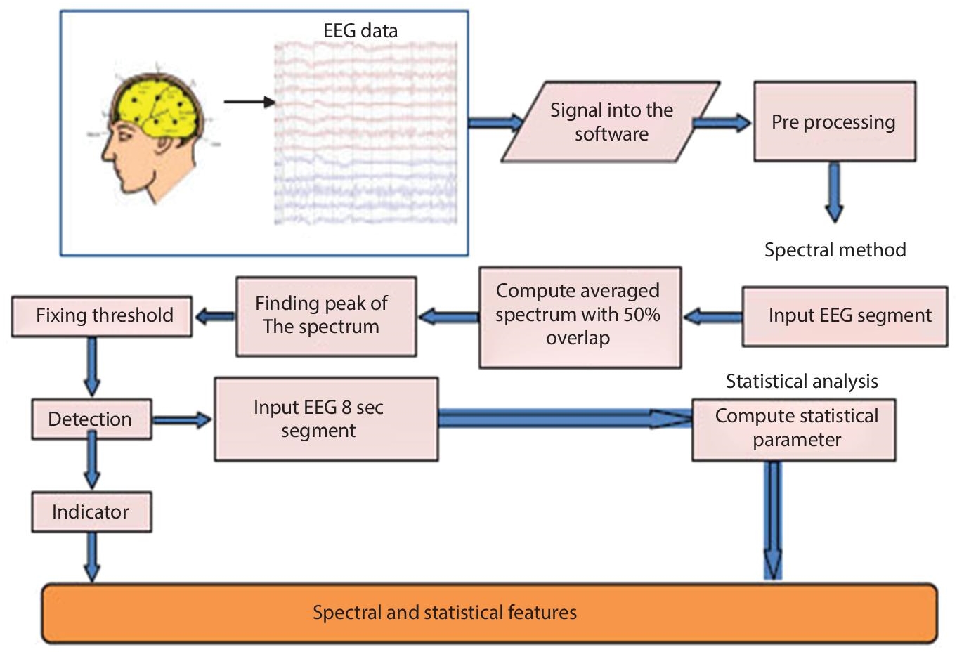
Figure 7.3 Design of block diagram for seizure detection algorithm.
The issue of characterizing the sign as being from an epileptic source is planned to grouping RPS pictures (Figure 7.4 shows the abnormal electrical brain activity due to seizures) of epileptic/non-epileptic subjects utilizing profound learning. Preparing profound learning models requires an enormous dataset of pictures and various emphases for convergence. As getting a huge dataset is testing, we embrace move learning for the task. Move learning plans to utilize the information gained from the source area to the objective spaces. It empowers us to make exact models without preparing a whole organization without any preparation even with a deficient dataset. Instead of gaining without any preparation, we start with designs realized when taking care of an alternate issue. Accordingly, we acquire information (highlights and loads) from recently educated models to prepare new models and try not to continue from scratch. Pre-prepared CNN models like LeNet, AlexNet, VGGNet, GoogLeNet, ResNet, and so on, can be utilized for the assignment.

Figure 7.4 Epileptic seizure.
In this investigation, a pre-prepared AlexNet model is utilized as it shows better execution in ordering epileptic seizure sets when contrasted with another best in class of CNN models to be specific LeNet and GoogLeNet. AlexNet changed every one of the records of prior non-profound learning-based strategies. AlexNet contains five convolution (Conv) layers and 3 completely associated (FC) layers. Every convolution layer comprises of 96–384 channels and the size of the channels goes from 3×3 to 11×11 with the element guide of 3–256 channels each. In each layer, a non-direct ReLU (Rectified Linear Unit) enactment work is utilized. ReLU is a significant component of AlexNet rather than the tanh or sigmoid actuation work used to prepare a model for a neural organization. The essential purposes behind utilizing ReLU in Convolution layers are quicker union attributable to the absence of disappearing slope issue and inciting sparsity in the highlights. 3×3 Max pooling is applied to the yields of layer 1, 2 and 5. In the main layer, a step of 4 is utilized to lessen the calculation.
For preparing the proposed model, 90% of epileptic and non-epileptic RPS pictures of the dataset (segment 5.1) are used (75% preparing and 15% approval); 10% of the information is saved for testing. To enhance the presentation defined 10-crease cross approval (CV) is performed. The 10-overlay CV parts the information at irregular into 10-disjoint sub-sets called folds. The defined folds keep up the mean reaction esteem in all folds, which is roughly equivalent. Each crease holds similar extents of the two kinds of class names, in particular epileptic (Set E) and non-epileptic (Sets A, B, C, and D) classes.
Preparing AlexNet model requires weight (Kernels) to be gained from the information. We use back-propagation with cross entropy as the misfortune work alongside the stochastic angle drop for improvement to get familiar with these boundaries. The model is prepared for picture characterization utilizing the Caffe structure. This model is prepared for 200 ages with learning rate (0.01), clump size (128), weight rot (0.0001), gamma (0.1), and force (0.9) as hyper-boundaries. The result is that the model’s profundity is huge for its high productivity, which is computationally costly yet made conceivable utilizing designs handling units (GPUs). A few other convoluted CNNs can perform adequately on quicker GPUs, even on enormous datasets. Utilizing K80 GPU machine, this progression takes around 45 minutes for one run of preparing for calculation on UoB datasets.
In view of data from magnetoencephalogram, electromyogram, electrooculogram, electrocardiogram, and EEG, nonlinear unique procedures are successfully utilized in biomedical applications [2–8]. This examination centers around displaying the nonlinear elements of the cerebrum. As generally acknowledged, the mind is viewed as a tumultuous powerful framework, and it produces EEG flags that are typically chaotic [9]. In another sense, an EEG signal as shown in Figure 7.5 is turbulent, as its sufficiency changes haphazardly after some time. These turbulent signs are described by long haul capriciousness, which makes traditional sign preparing strategies less accommodating. Demonstrating the elements is a test when utilizing regular highlights/models.
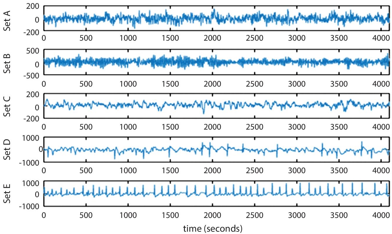
Figure 7.5 Seizure detection.
The AUC results for a couple of classifiers with mean error (as shown in Table 7.1) also showed improvements, with the KNNC classifier achieving 93%. An example for seizure classifier is given in the Figure 7.6. Given that sensitivities are more essential than specificities in this study, this is empowering. The underlying necessity is to gather the dataset of cerebrum signals. Table 7.2 shows the known seizure detection types and their acronyms. For this, diverse observing apparatuses are utilized. EEG and ECoG are the most often used devices since in channels or anodes were embedded to stick on the outside of scalp accordingly at range of 10–20 Global frameworks in various flaps. Every one of them has a wire association with the EEG gadget, giving convenient data about the varieties in voltage, alongside worldly and spatial data.
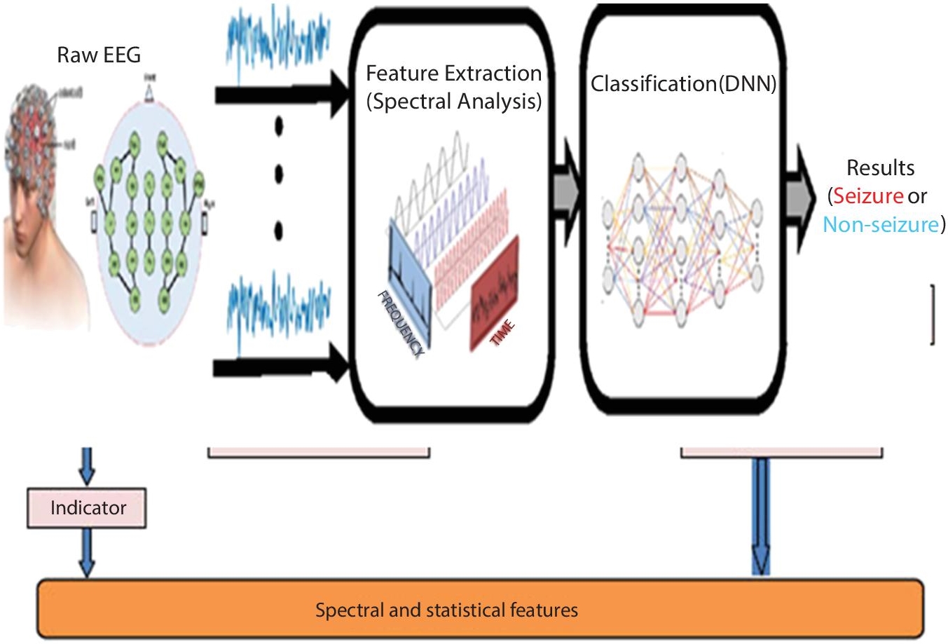
Figure 7.6 DNN-based seizure classifier.
Table 7.2 Acronym of seizure detection.
| S. no. | Acronym | Type of detection | Explanation |
|---|---|---|---|
| 1. | TP | True positive | Seizure detected |
| 2. | TN | True negative | Non-seizure (no disorder) detected |
| 3. | FP | False positive | Wrong detection of normal patient as seizure |
| 4. | FN | False negative | Inaccurately detecting seizure patient as normal |
There are three parameters for measuring seizure. They are precision, recall, and F-Measure. The ration of true positive to overall positives observed is known as precision. The percentage of true positive events is referred to as recall. The choral precision and recalls the estimated using the F-measure. For information researchers and scientists, a collection of data utilized to significant designed for assessing the presentation of others proposed models. We ought to capture cerebrum signals in epileptic seizure detection. EEG monitoring the most common way intended observing for brain activity. These accounts play a key in AI classifiers that look at novel seizure detection techniques several ways, as early capture diagnosis, quick seizure venue, enduring confiscation exploration, and annexation limitation. Value of publicity is available in the datasets of a baseline to compare and contrast results. Well-known collections of data are widely used in epilepsy will be depicted in the adjacent region.
It is discovered that contrasted with seizure identification, seizure confinement hasn’t seen much use of AI classifiers. However, there is some writing on the topic. In these instances, detailed work, creators didn’t specify the level of the influenced locale of the mind by a seizure and they couldn’t distinguish the specific area at the flaps like occipital, front facing, parietal left and parietal right. Despite the fact that, it isn’t our essential goal in this survey paper, while examining the connected distributed exploration, we tracked down some fascinating signs for seizure restriction.
7.3 Experimental Results
The critical factual highlights were extricated by various sorts of change procedures; Discrete Wavelet Changes (DWT), Consistent Wavelet Change (CWT), Fourier change (FT), Discrete Cosine Change (DCT), Solitary Worth Disintegration (SWD), Characteristic Mode Work (IMF), and time–recurrence space in EEG channel. These highlights guarantee to appropriate for capture recognition and in cerebrum linked applications like PC interface (BCI). Asserted exhibition stood assessed utilizing the accompanying measurements like affectability, explicitness, F-score, collector working attributes (ROC) bend, and percentile bootstrap measures.
This dataset involved EEG accounts got from five pharmacoresistant worldly flap patients with 3750 central and non-central bi-variant EEG records. Three affected roles were without seizure, with two patients just taking atmospheres yet no different seizures following a medical procedure. An intracranial strip and profundity cathodes were used to capture the multiple channel EEG signals as shown in Figure 7.7. The terminals were implanted in 10–20 positioning. Depending on if the EEG signals were registered with more than 64 channels, they were measured at 512 or 1024 Hz.
Utilizing the CNN structure, RMSprop (5) calculation and EEG signal, the learning of the weight boundaries of CNN was performed. Keras, an incredible profound learning library that sudden spikes in demand for top of TensorFlow, was used for displaying. During preparing, the cluster size for the information was chosen as 100, and this was used for every one of the preparation refreshes. Since the info information is a one-dimensional cerebrum signal, the CNN model is intended to acknowledge one-dimensional information of size 4096. Z-score standardization was applied for the scaling of each info signal and the upgrade of model speculation was figured it out. Subsequent to getting the info information, the first convolutional activity was applied to the info information and afterward the second convolutional activity was applied to the consequence of the first convolutional activity. Subsequently, the pooling was applied to the yields of the convolutional layer. Pooling diminishes the elements of the information.
Expanding the quantity of convolutional layers will permit us to acquire all the more profound highlights; however, this likewise builds the computational time. The completely associated network with three layers is applied for grouping purposes.
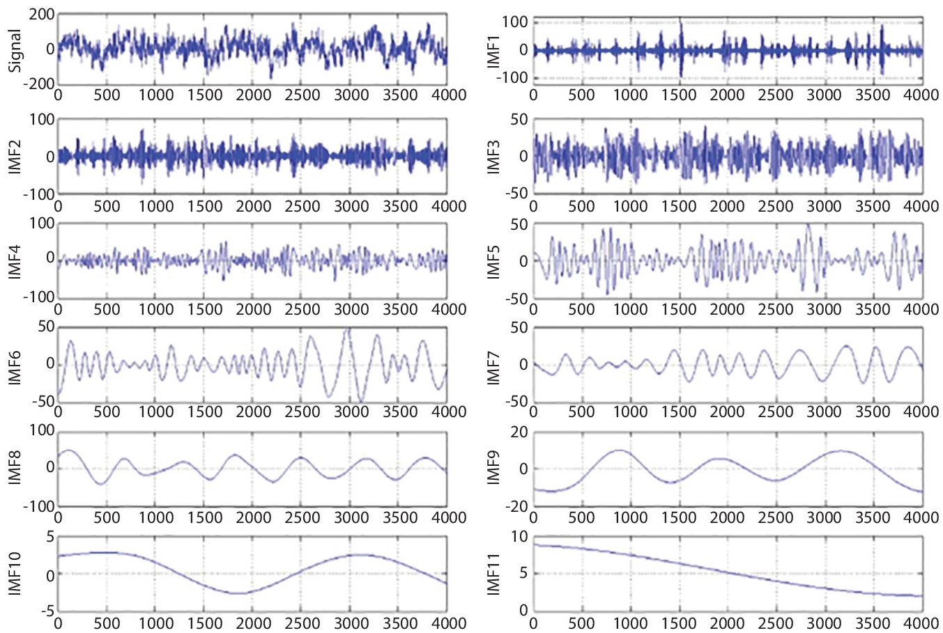
Figure 7.7 Simulated EEG signals with epilepsy.
We performed weight regularization to improve the learning of the CNN. This permitted us to decrease over-fitting prompting quicker advancement of the CNN model. We likewise rearranged the information previously parting it into the preparation and testing sets. Around there, the classes were similarly disseminated over the preparing and testing sets. Then, we applied the dropout activity. A profound neural organization needs to get familiar with countless boundaries, which on account of a little dataset is probably going to cause over-fitting. This issue is addressed by planning dropout innovation to forestall include locators from co-adapting. The vital idea of dropout is to drop units haphazardly from the neural organization during preparing, with a predefined likelihood (alongside their associations). Dropout strategy significantly takes out over-fitting and gives significant benefits over different types of regularization. We presented the dropout layer in the proposed model after the last ReLU actuation work. The built model incorporates include extraction and characterization modules, which improves the design of the epilepsy ID model. By including more convolutional layers or utilizing other non-direct capacities, we can expand the exhibition qualities and furthermore the precision of the model. Be that as it may, this will prompt complexity of the design and learning interaction of the model. Subsequently, diminishing the quantity of layers prompts an abatement in the precision pace of CNN.
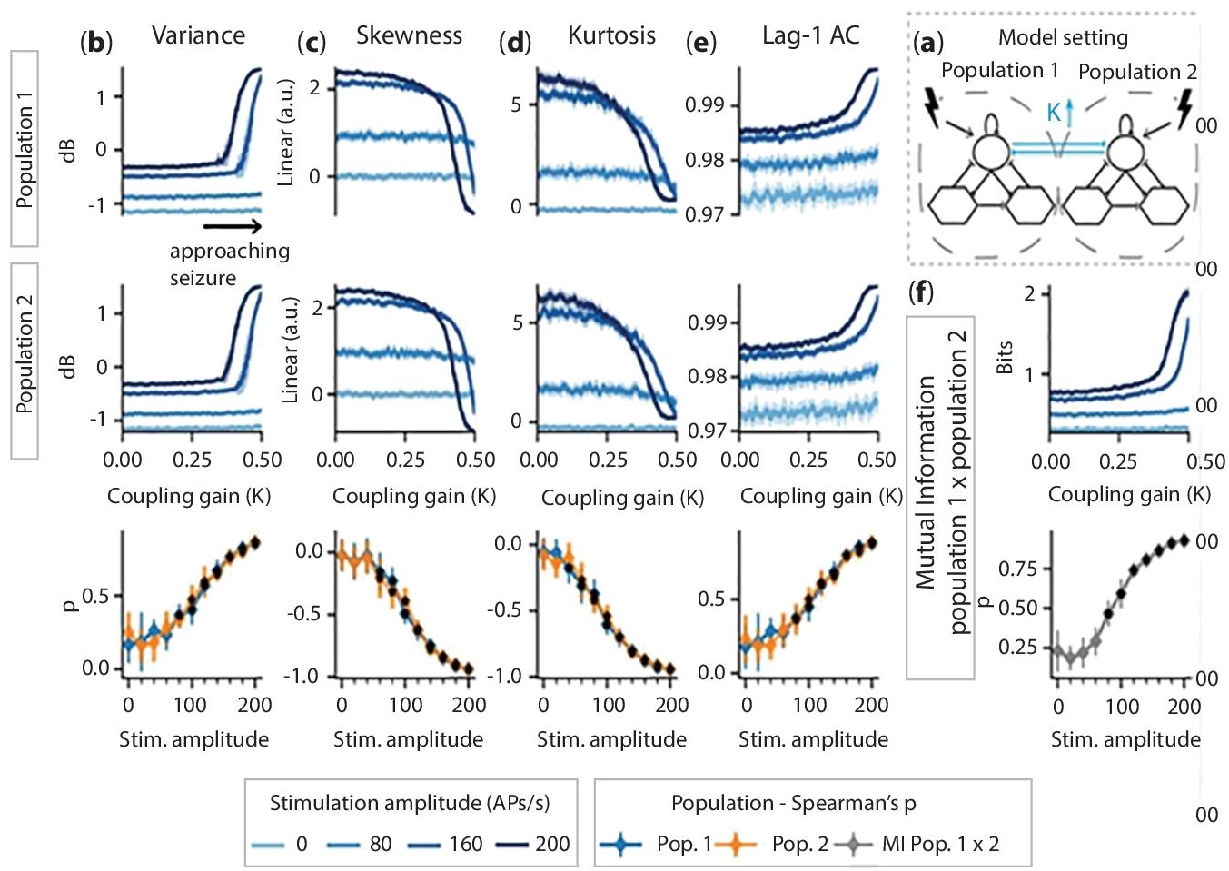
Figure 7.8 Experimental results of deep learning model based seizure detection.
Some measurable highlights were separated from EEG signals and these highlights were used as contribution to a strategic model tree (LMT) for epileptic seizure ID. The introduced technique was tried utilizing benchmark EEG dataset. The detection results are given in Figure 7.8. The papers referenced above portray various philosophies utilized for extraction of highlights and grouping purposes. The correctness’s of these planned models are significant execution attributes of the planned frameworks. In the paper [21] that utilized discrete wavelet change (DWT), the EEG signals were divided into recurrence sub-groups prompting the extraction of factual highlights. The PCA, free segments examination (ICA) and straight discriminant investigation (LDA) were used for information size decrease.
7.4 Taking Care of Children with Seizure Disorders
Managing a seizure condition in a kid relies on a variety of circumstances, including the age, overall health and type of seizure that occurs. Seizures in children can be treated.
Seizure medications: For children with seizure disorders, a wide range of medicines can be administered. Doctors do tests such as blood work, urinalysis, and EEGs to check how patients are reacting for a particular drug and if required, modify the type of medication.
7.5 Ketogenic Diet
A low-carbohydrate, high-protein, high-fat diet and induce the body to generate ketones, energy when carbohydrate intake is insufficient. The generation of ketones is critical to the effectiveness of this diet in terms of seizure management. The ketogenic diet should only be tried under the supervision of a physician.
7.6 Vagus Nerve Stimulation (VNS)
Process that involves surgically implanting a tiny battery into the chest and connecting it to the vague nerve using thin cables (located in the neck). When a kid detects the onset of a seizure, a magnet is placed over the battery, will stop the seizure.
7.7 Brain Surgeries
Brain surgery may be a possibility if a child’s seizures are uncontrollable by conventional means. This entails either eliminating the portion of the brain that is generating the seizures or blocking the propagation of seizure-inducing electric currents. While considering the brain surgery for seizures, a vital to think about how the removal of seizure-causing ports could influence other processes.
Consult experienced medical specialists to identify the best treatment choices for your kid. Individual symptoms will determine a safe and effective seizure therapy choice.
Some want to have a seizure dog in addition to medical assistance. These animals are taught to notify family members and shield the kid from danger during uncontrollable movements, or activate an alarm system in the event of a seizure. They can even predict seizures before they happen in some situations.
7.8 Conclusion
With the expansion of epilepsy, its precise identification turns out to be progressively significant. A significant test is to recognize seizures accurately from an enormous dimension of information. Due to intricacy of EEG signals in several datasets, AI classifiers suitable for precise detection of seizure. Regardless, picking out the suitable classifiers and highlights is, critical. Thus, this paper has exhaustively checked on AI approaches for seizure discovery. Accordingly, we presume that ‘non-discovery’ classifiers— choice woodland (outfit of choice trees) is best. This is on the grounds that it can deliver numerous reasonable, illustrative logic rules for forecasting with high accuracy. Further, it may also assist in the discovery of relevant information such as seizure restriction and seizure forms. Actually, ‘discovery’ classifiers can’t produce rationale rules, despite the fact that they can accomplish high prescient precision. Concerning choosing appropriate highlights, we should choose those that can give consistent outcomes. All the patients have been tested and were encountered up to 2–5 seizures, and a dataset consists of chronicles of 87 seizures from 21 patients. In the view of data collections, 6 contacts has been chosen for better understanding through visual review in the iEEG information by knowledgeable epileptologists: three close to the epileptic center (epileptogenic zone) and other three were in far off areas associated with annexation spread and proliferation. The subject went in age from the range of 10–50 years and infused with 13 ladies and eight men. Three distinctive type of seizure was addressed mid of the subjects, with basic incomplete (SP), complex halfway (CP), summed up tonic-clonic (GTC), and all subjects obligated encountered in any event two sorts. The epileptic center was situated in neocortical mind edifices with eleven patients, then hippocampus in eight patients, and two patients in two areas. The annexation begins occasionally and epileptic form exercises were commented by the ensured epileptologists at the Epilepsy Centre.
The characterization of interictal, ictal and preictal signals, just in precision of each tolerant was introduced; normal exactness of grouping among 21 patients was 92.3%. Patients were correctness’s order for nine patients with >95%, which has been viewed as an incredible outcome, and the characterization exactness was useful for eight patients, with values going somewhere in the range of 90% and 95%. The precision of sign grouping for the other four patients was <90%.
Seizure expectation is characterized as the programmed acknowledgment of forthcoming seizures where the forecast window can be up to a few minutes [18]. Achievement in foreseeing epileptic seizures would do horde advantages such as evasion of wounds, calming nervousness, crisis help and early mediation (for example early drug, electric incitement of vague nerve, profound cerebrum incitement). Four key stages are basic to investigation of seizure expectation. They are: 1) “interictal”, demonstrating the “ordinary” cerebrum state which is a long way from seizures; 2) “preictal “alluding to the time stretch preceding the seizure; 3) “ictal”, as time-frame in which seizure0happens; 4) “postictal stage”, relating to the period following a seizure and before an “ordinary” mind state. The preictal period, described by powerful development of EEG signals preceding seizures is the examination center for the seizure expectation. Because of the powerful idea of preictal EEG, it is accounted for that the hypothesis of turbulent elements exhibits preferred unsurprising capacity over direct estimations [13]. The benefits of bi-variate and multivariate gauges over uni-variate (for example ghastly force) have been demonstrated [14].
Finding the essential epileptogenic zone is troublesome, since electric movement of seizure may upheaval unexpectedly and all the while proliferating over a wide scope of cortical zones. Signs gathered by intracranial cathodes with high goal are viewed as the ideal to research the SOZ. Among them, high recurrence swaying (HFO) conveys data distinction from low-recurrence releases, which is featured as SOZ corresponded bio-markers in epilepsy. In the past examinations, a few basic highlights or segments (quick movement, signal leveling, moderate expected shift, and so forth) have been investigated utilizing intracranial anodes (counting stereotactically-embedded intracranial EEG [SEEG], subdural electrocorticography [ECoG], and so forth) Given seizure beginning is a perplexing marvel which is created by different spatio-worldly components, focusing on single element can’t be assessed in disconnection. Consequently, we construed ML procedures could address those worries. Grinenko et al. built up a SVM-based learning model to find a “unique mark”, which adequately separated time-recurrence examples of the SOZ from spaces of engendering [24].
ML as an arising method renders principled, programmed and target calculations for high-dimensioned furthermore, convoluted information, which shows its benefit in EEG signal investigation contrasted with conventional techniques. Regardless of the benefits of ML (for example little biasness and high affectability to designs acknowledgment), solid classifiers, explicit component extraction, very much chose information and calculation cost should all be thought about. For instance, channel choice on specific conditions can decrease calculation stacking of both element extraction and example acknowledgment, which is attainable for the on-line calculation particularly for some wearable or implantable gadgets in genuine application. Another likely option is to exploit distributed computing connected by 5G innovation to acknowledge constant trade of EEG recording. As for ML techniques, profound learning as a hot procedure in picture preparing, yet it is simply beginning to arise in EEG handling, which show its predominance in EEG design acknowledgment
References
- 1. Tzimourta, K.D., Tzallas, A.T., Giannakeas, N., Astrakas, L.G., Tsalikakis, D.G., Angelidis, P., Tsipouras, M.G., A robust methodology for classification of epileptic seizures in eeg signals. Health Technol., 9, 2, 135–142, 2019.
- 2. Alickovic, E., Kevric, J., Subasi, A., Performance evaluation of empirical mode decomposition, discrete wavelet transform, and wavelet packed decomposition for automated epileptic seizure detection and prediction. Biomed. Sign Process. Contr., 39, 94–102, 2018.
- 3. Shimizu, M., Iiya, M., Fujii, H., Kimura, S., Suzuki, M., Nishizaki, M., Left ventricular end-systolic contractile entropy can predict cardiac prognosis in patients with complete left bundle branch block. J. Nucl. Cardiol., 3, 1–10, 2019.
- 4. Altunay, S., Telatar, Z., Erogul, O., Epileptic EEG detection using the linear prediction error energy. Exp. Syst. Appl., 37, 8, 5661–5665, 2010.
- 5. Lee, S.-H., Lim, J.S., Kim, J.-K., Yang, J., Lee, Y., Classification of normal and epileptic seizure eeg signals using wavelet transform, phase-space reconstruction, and Euclidean distance. Comput. Methods Progr. Biomed., 116, 1, 10–25, 2014.
- 6. Chandaka, S., Chatterjee, A., Munshi, S., Cross-correlation aided support vector machine classifier for classification of EEG signals. Exp. Syst. Appl., 36, 2, 1329–1336, 2009.
- 7. Moslem, B., Karlsson, B., Diab, M.O., Khalil, M., Marque, C., Classification performance of the frequency-related parameters derived from uterine EMG signals, in: Proceedings of the International Conference of the IEEE Engineering in Medicine and Biology Society, Massachusetts, USA, Boston, pp. 3371–3374, 2011.
- 8. Hassan, M., Terrien, J., Marque, C., Karlsson, B., Comparison between approximate entropy, correntropy and time reversibility: Application to uterine electromyogram signals. Med. Eng. Phys., 33, 8, 980–986, 2011.
- 9. Wang, C.-M., Zhang, C.-M., Zou, J.-Z., Zhang, J., Performance evaluation for epileptic electroencephalogram (EEG) detection by using Neyman–Pearson criteria and a support vector machine. Physica A: Stat. Mech. Appl., 391, 4, 1602–1609, 2012.
- 10. Patnaik, L.M. and Manyam, O.K., Epileptic EEG detection using neural networks and post-classification. Comput. Method. Prog. Biomed., 91, 2, 100– 109, 2008.
- 11. Srinivasan, V., Eswaran, C., Sriraam, N., Approximate entropy-based epileptic EEG detection using artificial neural networks. IEEE Trans. Inform. Technol. Biomed., 11, 3, 288–295, 2007.
- 12. Panda, R., Khobragade, P.S., Jambhule, P.D., Jengthe, S.N., Pal, P.R., Ghandhi, T.K., Classification of EEG signal using wavelet transform and support vector machine for epileptic seizure diction, in: Proceedings of the International Conference on Systems in Medicine and Biology, Kharagpur, India, pp. 405– 408, December 2010.
- 13. Nasehi, S. and Pourghassem, H., Patient-specific epileptic seizure onset detection algorithm based on spectral features and IPSONN classifier, in: Proceedings of the 3rd International Conference on Communication Systems and Network Technologies (CSNT ‘13), Gwalior, India, IEEE, pp. 186–190, 2013.
- 14. Williamson, J.R., Bliss, D.W., Browne, D.W., Epileptic seizure prediction using the spatiotemporal correlation structure of intracranial EEG, in: Proceedings of the IEEE International Conference on Acoustics, Speech, and Signal Processing (ICASSP ‘11), Prague, Czech Republic, IEEE, pp. 665–668, May 2011.
- 15. Taft, L.M., Evans, R.S., Shyu, C.R. et al., Countering imbalanced datasets to improve adverse drug event predictive models in labor and delivery. J. Biomed. Inform., 42, 2, 356–364, 2009.
- 16. Chandaka, S., Chatterjee, A., Munshi, S., Cross-correlation aided support vector machine classifier for classification of EEG signals. Exp. Syst. Appl., 36, 2, 1329–1336, 2009.
- 17. Polat, K. and Gunes, S., A novel data reduction method: Distance based data reduction and its application to classification of epileptiform EEG signals. Appl. Math. Comput., 200, 1, 10–27, 2008.
- 18. Khan, Y.U., Rafiuddin, N., Farooq, O., Automated seizure detection in scalp EEG using multiple wavelet scales, in: Proceedings of the IEEE International Conference on Signal Processing, Computing and Control (ISPCC ‘12), IEEE, Waknaghat, India, pp. 1–5, March 2012.
- 19. Chandaka, S., Chatterjee, A., Munshi, S., Cross-correlation aided support vector machine classifier for classification of EEG signals. Exp. Syst. Appl., 36, 2, 1329–1336, 2009.
- 20. Wang, Y., Simon, M., Bonde, P. et al., Prognosis of right ventricular failure in patients with left ventricular assist device based on decision tree with SMOTE. IEEE Trans. Inform. Technol. Biomed., 16, 3, 383– 390, 2012.
- 21. Acir, N. and Guzelis, C., Automatic spike detection in EEG by a two-stage procedure based on support vector machines. Comput. Biol. Med., 34, 7, 561–575, 2004.
- 22. Leman, H., Marque, C., Gondry, J., Use of the electro-hysterogram signal for characterization of contractions during pregnancy. IEEE Trans. Biomed. Eng., 46, 10, 1222–1229, 1999.
- 23. LeVan, P., Urrestarazu, E., Gotman, J., A system for automatic artifact removal in ictal scalp EEG based on independent component analysis and Bayesian classification. Clin. Neurophysiol., 117, 4, 912–927, 2006.
- 24. Blanco, S., Kochen, S., Rosso, O.A., Salgado, P., Applying time-frequency analysis to seizure EEG activity. IEEE Eng. Med. Biol. Mag., 16, 1, 64–71, 1997.
Note
- *Corresponding author: [email protected]