
Chapter 10
Identifying Unknown Victims
IN THIS CHAPTER
![]() Outlining how bodies are identified
Outlining how bodies are identified
![]() Finding answers in skeletal remains
Finding answers in skeletal remains
![]() Determining the time and cause of death
Determining the time and cause of death
![]() Using facial reconstruction and photographic techniques
Using facial reconstruction and photographic techniques
Not every corpse that comes into the coroner’s office is conveniently carrying a driver’s license and Social Security card. All too often police are confronted with identifying an unknown corpse. In movies, this process usually takes only a few minutes of screen time, but in reality, it may take weeks, months, or even years. Some bodies never are identified.
The corpse in question may have been dead for hours, days, months, years, or many decades. An extended time since death and many other factors can complicate the identification process, which usually involves several different forensic disciplines and techniques, all of which are coordinated by the medical examiner (ME).
Identifying the Body
Neither Mother Nature nor time is kind to the dead. Not only is a dead body subjected to internal digestion (autolysis) and bacterial action (putrefaction), but extreme weather, insects, predators, and environmental bacteria conspire to destroy it.
 The condition of a body when it’s found depends on how long ago the death occurred and whether it is left exposed to the elements or buried. The general rule: One week in the open equals two weeks in water or eight weeks in the ground. The more decomposed the body, the greater the medical examiner’s challenge in making an ID.
If the body is more or less intact, its size, sex, race, scars and tattoos, facial features, dental examinations, fingerprints, DNA, and clothing may help authorities identify who it is. So an intact or partially decayed body gives the ME a great deal to work with and in many cases, a facial photograph of the corpse can be taken and compared with photos or descriptions of missing persons. Problem solved. However, when the body is skeletal, help from a forensic anthropologist, forensic odontologist (dentist), forensic artist, or all three may be needed.
Once a presumptive (possible or likely) match is made, family or friends may be asked to make the final identification. If no missing person matches the general characteristics of the corpse, descriptions and photos are circulated to law enforcement agencies and the media. To help in this regard, a forensic artist or anthropologist may be asked to sketch or computer-generate a best-guess likeness of the individual (more on this in the later section “Reconstructing Faces”).
Sifting through the artifacts
Sometimes items buried with or found near the body are helpful. Clothing, jewelry, materials used in the burial, and other artifacts can point to who the deceased is. Finding a wallet or ID card is ideal, but even if that sort of information is found, the forensic team may be involved in the ID process if the person cannot be positively identified visually. Many other items, however, offer clues:
- Rings or jewelry often are inscribed with names, initials, or dates.
- Clothing may be distinctive in either style or manufacturer. For example, designer clothes and shoes can point the police in one direction, while ragged and worn clothes like those worn by a homeless person point them in another.
- A coffin, blanket, or some other material may have been used to bury the victim by a criminal hoping to keep animals away and make the body harder to find.
- A makeshift, wooden coffin may provide information from its construction materials and methods as well as from any distinct markings it bears.
- Blankets or sheets likewise may bear tags identifying the manufacturer or seller.
- A plastic bag may offer up the perpetrator’s fingerprints, which, in turn, can lead to the identity of the corpse.
Examining scars, birthmarks, and tattoos
Distinguishing marks on the body, such as tattoos, birthmarks, and surgical or other scarring, often are helpful in suspect and corpse identification. Many are so distinctive that they alone supply positive identifying evidence. Could you mistake former Soviet President Mikhail Gorbachev for anyone else? Probably not, because the port wine stain on his noggin makes his face one of a kind.
Birthmarks are irregular and distinctive, making them perfect identifying characteristics. The tattoos and other body marks of arrested criminal suspects often are sketched or photographed as part of the booking process; however, this procedure is far from universal. If photos from a previous arrest exist, they can be compared with the body marks on a suspect or corpse.
 Tattoos, even when they’re not readily identifiable, may be traced to the artist by style, chemical composition, or both. Many tattoo artists use black pigments that contain carbon, reds that contain mercuric chloride, and greens that contain potassium dichromate. Others use aniline-based dyes. Extracting and analyzing some of the pigment from the tattooed skin of a corpse makes confirming or excluding the work of a specific tattoo artist possible and takes investigators one step closer to identifying the victim.
Certain gangs boast their own tattoos, and many jurisdictions keep files of these tattoos. In California, for example, the CALGANG database stores such data, and often a query results in a hit, which leads to identifying the victim. If the deceased had a previous brush with the law, a former cellmate, corrections officer, or arresting officer may be able to supply a presumptive ID.
Finding evidence of wounds or disease
During an autopsy, the ME may discover that the deceased suffered from a disease or sports a scar from a surgical procedure. This information narrows the search for identification. If the victim has a scar from an appendectomy or gall bladder removal, for example, a search through missing-person reports for people who are the same age and sex and have the same medical history may lead to identification, particularly if the surgery was a fairly recent event. The ME often can determine the age of surgical wounds.
 Any wound, regardless of whether it’s from surgery, a knife fight, or other means, follows the same healing pattern. During the first week, surgical and repaired knife wounds usually require sutures (or stitches). For several months following the removal of the sutures, a telltale pattern caused by the suturing can be seen. The pattern, however, may fade after several months.
For several weeks, any scar remains slightly pink to brownish-red because of microscopic blood vessels supplying blood to the area to aid in the healing process. During the next few months, the body continues to repair the damage by laying down collagen (thick strands of connective tissue), the color gradually fades, and the scar shrinks considerably. As scars mature, they finally become faint white lines after four to six months. The collagen layering continues to shrink over the next year or so. Thereafter, the scar remains pretty much the same. Thus, the age of a scar can be approximated within the first four to six months, but thereafter, all bets are off.
Certain surgical appliances provide another means of identification, because they are uniquely marked. The ME may discover through an X-ray exam or during the autopsy that the deceased had a hip replacement surgery, for example. The artificial hip is removed and examined for an engraved serial number, which then can be traced to the hospital where the surgery took place and ultimately to the name of the person who received it. Pacemakers, implantable defibrillators, heart valves, and other cardiac devices have traceable serial numbers.
Fingerprinting the dead
Unless a corpse is severely deteriorated, fingerprints can be obtained and matched against known missing persons and national fingerprint databases. Fingerprints often lead to quick and absolute identifications.
Saline sometimes is injected into the tips of the fingers, causing the pads to swell and potentially reveal the friction ridges (see Chapter 5). Alternatively, skin over the pads of the fingers can be carefully sliced away, viewed under a microscope, and even photographed for matching purposes.
 Fingerprints can even be obtained from mummified bodies in some circumstances. The finger pads of such corpses are shriveled and have the texture of old leather, but soaking them in water or glycerin may cause them to swell enough for fingerprints to be obtained.
Checking out the choppers
Everyone’s teeth are different. Although you and I may have the same number and types of teeth, their lengths, widths, and shapes show great variability. Missing, misaligned, and reconstructed teeth can be matched with dental records to establish an identity. Chips, furrows, and fillings add even more individuality to the patterns of your teeth. Today, dental records often are used to identify human remains. DNA also plays a role here as the pulp of the teeth often supplies DNA-containing tissues.
 Using teeth as a method of identity isn’t new. In the first century A.d., the Roman Emperor Claudius demanded to see the teeth of his beheaded mistress to assure her identity. He knew she had a discolored front tooth. William the Conqueror used his crooked teeth to bite the wax seals on his letters to prove they came from him. The first time teeth were used to identify victims of a mass disaster was after the 1849 fire at the Vienna Opera House.
When faced with an unidentified corpse, the ME often makes a set of dental X-rays that can be compared with the most recent dental X-rays of a missing person. With a match, the identity of the body is confirmed.
The ME uses any and all evidence she can glean from a corpse in order to ID the deceased. Even stomach contents can be of use in rare instances.
Using DNA To Make the ID
DNA is also used in identifying unknown remains in many cases. But, like fingerprints, it is only useful if there is a DNA profile available against which it can be compared. DNA taken from the corpse’s tissues, or from bones and teeth in the case of skeletal remains, can often be analyzed and compared to DNA from a missing person — if it’s available. It can also be entered into DNA databases such a CODIS in the hopes of getting a “hit” — a match with someone who is already in the system.
Besides nuclear DNA, other DNA analyses, such as mitochondrial DNA, Y-chromosomal DNA, and familial DNA, can be very useful in these situations as each can narrow the possibilities. Each of these techniques is discussed in Chapter 15.
Dem Bones, Dem Bones: Working with Skeletons
Sometimes a forensic team doesn’t have a complete body to work with. They may have only a skeleton (at best) or simply a single bone. In these situations, the expertise of a forensic anthropologist and a forensic odontologist usually come into play. They’re asked to answer several questions:
- Are the bones human?
- What are the biological characteristics (size, age, sex, and race) of the individual?
- How long has the person been dead?
- What is the cause and manner of death?
With an intact adult skeleton, determinations of sex can be made essentially 100 percent of the time, age to within 5 to 10 years, height to within 1.5 inches, and race much of the time. The problem: Often only a portion of the skeleton or only a few bones are available to the anthropologist.
Any hope of identifying an unknown corpse rests with determining the deceased individual’s biological characteristics; age, stature, sex, and race narrow the field greatly. After these characteristics are determined, the search turns toward individualizing characteristics. The presence of bony evidence of disease, congenital defects, or trauma is important.
Determining whether bones are human
The first question that must be answered is whether the bones are human. Time and the effects of nature and predators can scatter and destroy portions of a skeleton, leaving investigators with little to go on and no room for assumptions.
Determining the species (let alone the specific identity) of a bone is challenging, to say the least. The front paw bones of a bear are eerily similar to those of a human hand, for example, and shell fragments from some turtles can be mistaken for skull fragments. Ribs from sheep and deer resemble human ribs. If the victim is an infant or young child, determining that the bones are human can be even more difficult. Infant bones are much smaller and easier to confuse with the bones of small animals. An infant’s skull is not fully fused (joined together into a single structure) and some of these fragments can be scattered and lost, so a complete view of the skull is not apparent. Infant teeth also resemble those of small animals.
Bones are more complex structures than you may realize. They have bumps, grooves, indentations, and other characteristics according to their function in the body and what species that body belongs to. The forensic anthropologist uses these features, as well as overall size and thickness, to assess the species of origin. Yet, even with years of experience, species identification can prove difficult.
Determining age
When only bones are available, the forensic anthropologist can only make a best guess (which is, after all, a scientific opinion) about age, but that estimate is more accurate for younger victims than it is for mature victims. Teeth and bones in children and adolescents follow a predictable growth and maturation pattern. By assessing the stage of this development, a fairly narrow age range can be determined. Later in life, after the maturation process is complete, changes in the teeth and skeleton occur at much slower rates, thus leading to age assessments across much wider ranges.
Details that are particularly useful include
- Teeth: Tooth development begins before birth and progresses through the formation, appearance, and loss of baby teeth (20 altogether) and ultimately the appearance of 32 permanent teeth. The appearance of permanent teeth is complete usually by about age 12. The last teeth to appear, the wisdom teeth, typically erupt by age 18. This general timeline helps with assessing the age of anyone who’s 18 or younger at the time of death.
- Skull: In adults, the skull is of little use for age estimation, and in infants it can be of some help, but not as much as once believed. The skulls of infants actually are in several pieces. With time the pieces fuse or meld together along jagged lines of separation known as suture lines. Although it seems logical to conclude that the pattern of the closure of the suture lines is useful, unfortunately suture line fusion occurs in such a widely variable pattern that age estimation is not as accurate as investigators would wish.
- Long bones of the legs and arms: These bones change as the body ages. The growth plates within them remain open as they grow, but then close when growth comes to an end. These changes can help determine the ages of people younger than 25, when the bones have completed their growth.
- Pelvis: The symphysis, a thin band of cartilage, fuses together the front of your pelvis. It has a zigzag shape in the beginning, but it straightens as you age, stopping when you reach about 50.
- Ribs: Areas where ribs join the breastbone, or sternum, are smooth and rounded when you’re young, but they become pitted and sharp as you age. Examination of these junctions can narrow age prediction to within 1 and 1/2 years up to age 30 and within 5 years up to age 70. After that, these changes are of little use.
- Bone density: As you age, your bones lose calcium and become less dense. X-ray examination of the bones reveals the density of the calcium and may help with determining age. Malnutrition and diseases such as osteoporosis lessen the bone density at any given age, so those factors must be considered when determining age from skeletal remains.
Estimating stature
You may think that simply measuring the skeleton from top to bottom provides its height and general build. When a complete skeleton is present, that’s entirely possible; however, more often only partial remains are found, making such measurements out of the question. Remember that height is but one part of your stature, which also includes body shape, bone thickness, and degree of muscular development.
The long bones (legs and arms) can help provide an estimate of height. Tables matching length of long bones to the expected height of the person from which they came are available to help in this regard. For example, height usually is equal to five times the length of the humerus (upper arm bone). Even fragments of long bones can be useful in determining stature. Other formulas have been developed to enable investigators to estimate bone length from fragments. Such estimates make possible a rough calculation of the person’s overall height.
The thickness of the bones gives investigators a rough idea of whether the person was of slight or muscular build, as thicker bones often indicate thicker muscles. The thickness may also reveal handedness, because right-handed people usually have thicker, stronger bones on their right side, and vice versa for southpaws.
Determining sex
Determining sex from skeletal remains of infants and children is more difficult than it is with adults because gender-specific changes in the skeleton don’t appear until puberty, after which male and female bones grow differently and begin to take on sex-identifying characteristics.
The overall size and bone thickness of the adult male skeleton usually is greater than that of the adult female. However, bone size and thickness is related to many things other than sex: Better nutrition and heavy physical activity lead to stronger bones regardless of sex. So the skeleton of a female who ate well and performed manual labor may look more like that of a male than the skeleton of a poorly nourished male who rarely worked physically.
Nevertheless, the thickness of certain areas of certain bones can be used to distinguish between males and females. In general, the diameters of the heads of the humerus (upper arm bone), the radius (lower arm bone on the thumb side), and the femur (upper leg bone) are larger in males.
The most reliable bones for determining sex are those of the pelvis (see Figure 10-1). The male pelvis is designed only for support and movement, while the female pelvis is adapted for childbirth. The female pelvis is wider and has a wider pelvic outlet, which allows passage of the infant during childbirth. The sciatic notch (which the sciatic and other nerves pass through on their way to the leg) is wider in females than it is in males. In addition, the backside of the pubic bone of a woman who’s delivered a child may have pregnancy pitting, or scarring and irregularities caused by the tearing and regrowth of ligaments that occur during and after childbirth.
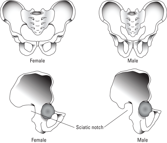
Illustration by Nan Owen
FIGURE 10-1: The female pelvis is wider than the male pelvis, and the sciatic notch is broader in females than in males.
The skull also offers useful clues to the sex of an individual. Male skulls tend to have more distinct ridges and crests and to be larger and thicker, particularly in areas where facial and jaw muscles attach. In addition, the posterior ramus (back branch) of the mandible (jaw bone) in males is slightly curved, but in females it tends to be straight (see Figure 10-2).
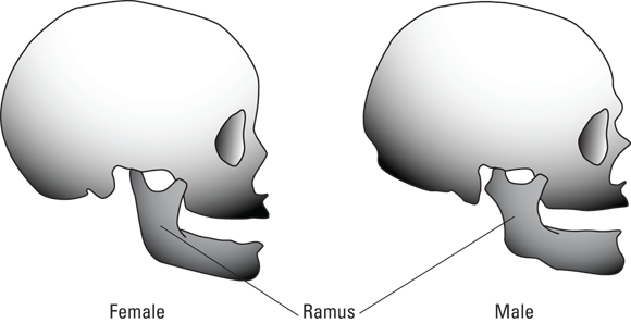
Illustration by Nan Own
FIGURE 10-2: The posterior ramus of the mandible is curved in males but straight in females.
Determining race
Using skeletal remains to determine race is extremely difficult, if not impossible, because no single skeletal trait is racially distinct. Any classification is rough at best and is greatly altered by racial admixture. The only three groups to which a given skeleton can be assigned are Caucasoid, Negroid, or Mongoloid.
Caucasians tend to have high, rounded, or square skulls, straight faces, and narrow, protruding noses. The shape of the eye sockets is triangular. On the other hand, those of Negroid descent tend toward lower and narrower skulls and wider, flatter noses with prominent, protruding teeth. Their eye sockets are usually squared. Mongoloids have broad, round skulls with an arched profile. The eye sockets are round with wide facial dimensions. Blacks tend to have proportionally longer arms and legs than do Caucasians, while in Mongoloids, the limbs tend to be shorter. Caucasians also have a forward curve to their femurs (upper leg bones), while in blacks this bone is straighter.
The skeleton of someone with a mixed racial origin obviously shares the skeletal characteristics of its ancestors. This racial admixture frequently makes racial determinations impossible.
Seeking individual characteristics
Estimates of the age, stature, sex, and race of the remains greatly narrow the search for the identity of the unknown person, but establishing the true identity requires much more information. Just knowing that the remains are those of a 12- to 18-year-old, 5-foot-tall, left-handed Caucasian female doesn’t absolutely identify the individual. Far from it. But these factors narrow the focus to a manageable number of individuals.
Other details that can be compared with medical records or X-rays of potential matches to make identification more certain include
- Evidence of previous injuries, such as healed fractures
- Evidence of knife or gunshot wounds, which can fracture and nick bones
- Surgical appliances, such as artificial hips and pacemakers
- Skeletal evidence of disease, such as the characteristics of bone cancers, tuberculosis, or rickets
- Dental patterns
- DNA extracted from bones or teeth
One newer technique that is being used for identifying remains is matching mitochondrial DNA (mtDNA — see Chapter 15) from a corpse with that of a survivor. Mitochondrial DNA is inherited and unchanged along maternal lines for many centuries. It is very hardy, survives for long periods of time in decayed and skeletal remains, and is found in all the cells of the body, the pulp of the teeth, and even in shafts of hair, which don’t contain normal, or nuclear, DNA. If the victim is believed to be a particular individual, then the mtDNA obtained from close relatives can be compared with that of the deceased person. If they share a common maternal ancestry, the mtDNA from each will match. Other useful DNA techniques include Y-chromosomal DNA and familial DNA analyses.
Estimating time since death
Taphonomy is the study of what happens to the human body after death. How the body decays, if it does, and how it becomes skeletonized (deteriorated to the point that only bones remain) are areas of interest to the student of taphonomy. As you’ve seen so far in this chapter and can discover in Chapter 11, estimating the time of death may involve not only the forensic anthropologist and odontologist, but also an ME, archeologist, climatologist, botanist, entomologist, and others (Chapter 2 gives details about each of these positions).
When confronted with skeletal remains, the forensic anthropologist first works to determine how old the bones are. The answer is critical to any forensic involvement. Bones that are hundreds of years old have little forensic use, but bones that are between 2 and 40 or so years old may have significant import.
Estimating the time since death is never easy, and it becomes increasingly more difficult with each passing hour. Chapter 11 looks at determining time of death of more-or-less intact corpses, a task that falls within the expertise of the ME. With skeletal remains, the forensic anthropologist determines the approximate time lapse since the death occurred, using any or all of the following methods:
- Examining artifacts from the burial site: Clothing, jewelry, casket materials, and burial artifacts may indicate the period of the burial. For example, arrowheads or musket balls suggest a different time frame than do synthetic materials and plastic objects.
-
Chemical analyses: Measuring the nitrogen level in bones, which decreases as they deteriorate, is one such method. This kind of analysis is inexact, however, because the rate of protein and nitrogen loss is affected by temperature and moisture, the same two factors that most affect the decay rate of the body. Nonetheless, because a high level of nitrogen suggests that the bones are a few years and not several decades old, this determination can be of some help.
Another chemical measurement is based on different amino acids disappearing from bones at different rates. Analysis of fresh bones may yield as many as 15 different amino acids, but bones that are a hundred or more years old often yield only seven.
- Ultraviolet (UV) light: Fresh bones fluoresce (glow) a pale blue color under UV light. A cross section of the bone reveals this glow across the full thickness of the bone. Time causes this fluorescence to diminish from the outside in, so a bone that is less than 100 years old may glow across its full thickness, whereas one that is several hundreds of years old shows no fluorescence at all. In between, the fluorescence may be confined to a narrow band within the bone’s cross section. This band narrows decade by decade until it disappears.
- Radioactive isotopes: Carbon-14 (C-14) dating is of little use in forensics because its ranges are too broad; however, other radioactive materials may be helpful. Continuous testing and use of nuclear weapons from World War II through the ’50s and ’60s caused global increases in the amounts of C14, strontium 90, cesium 137, and tritium (a radioisotope of hydrogen). Finding increased amounts of one or more of these substances in bones means that the person died after about 1950.
Handling burned bones
Bones that have been burned present special problems for the anthropologist. Direct exposure to the fire chars and blackens bones and may cause them to crack or splinter. Prolonged direct contact with fire can calcinate bones, reducing them to white ashes. Under those circumstances, an anthropologist may have only a few remnants to work with.
On the other hand, the texture and color of the bones provide clues about the intensity and the duration of the fire. Indirect or brief exposure to the fire may cause only yellow-brown discoloration of the bones with or without streaks of soot.
Desiccation, or drying out of the bones, causes them to shrink, thus making an accurate estimate of stature very difficult.
Determining cause and manner of death
Occasionally, skeletal remains offer clues to the cause and manner of death. Fractures, fragmentation, and impact marks (dents and depressions) on the bone may reveal blunt-force injuries, indicating the victim likely fell or was hit with a blunt object. Sharp-force injuries, such as from an axe or knife, may show up as cut surfaces where the blade sliced into the bone. Metallic remnants from the weapon occasionally remain along cut surfaces. Similarly, gunshots may leave entry and exit holes in the skull, and gouges and other defects in the ribs, spine, and other bones. Finding a bullet or two among the bones helps. Measuring an entrance hole in a bone sometimes enables investigators to estimate the caliber of the bullet that caused the injury.
Tracking injuries to more than one bone or finding an imbedded bullet may also enable investigators to estimate the path of the bullet and thus determine which organs may have been damaged. Of course, blunt objects, knives, and bullets can lead to death without impacting the skeleton, and, unfortunately, many strangulations and most natural deaths leave behind no skeletal evidence, meaning that a skeleton may not offer any clues to the cause and manner of death.
One problem facing the ME is whether the bone injuries occurred around the time of death or at some earlier point in time. For example, a skull fracture that occurred years before death and one that occurred at the time of death mean two entirely different things. Fortunately the forensics team often can make such a distinction.
Fractured bones heal, evidence of which can be seen by way of callus (scar) formation in the area of injury. Callus formation takes months to complete, and, of course, no healing occurs after death. So a fracture that sports a robust callus probably occurred months before death. Perimortem fractures (ones that occur close to the time of death), on the other hand, show no signs of healing and no callus formation. Thus, a skull fracture that shows no sign of healing may have occurred around the time of death and may, indeed, be related to the cause of death. Conversely, well-healed fractures can’t be directly related to the cause of death.
Bones left in nature undergo trauma from natural forces and from predators and can suffer fractures years after the victim’s death. Forensic anthropologists recognize postmortem fractures as such in large part because living bones possess moisture, living protein, and fat, and that makes them less brittle than bones from a long-dead victim.
Fractures to living bones tend to be spiral (twisting down the bone’s shaft) or greenstick (splintered, like when you snap a green twig in half). Desiccated (dried out) bones are brittle and tend to crumble more readily and break cleanly, usually parallel or at a cross section to the long axis of the bone. By examining the nature of any fractures, a forensic anthropologist may be able to distinguish premortem fractures from those that occurred postmortem.
Based on the timing and nature of skeletal injuries, forensic anthropologists and MEs sometimes can determine cause of death and perhaps even whether it was self-inflicted, accidental, or homicidal in nature. Determining the manner of death, however, is not always easy or accurate. When findings suggest that someone died from a skull fracture, determining whether the fracture was caused by a blow to the head (homicidal), a fall (accidental), or as the result of a fall brought on by a heart attack or stroke (natural) can be difficult.
Reconstructing Faces
Forget Michelangelo. For the poor victims who end up dead and unidentified, great artists are those who re-create faces based on skeletal remains. Whether through drawings, sculptures, or computer-generated images, these artists help identify a body by creating a likeness of the victim that is circulated in hopes of finding someone who can identify him. Facial reconstruction, or re-creating a likely image of the unidentified victim’s face, comes into play when other methods have failed to identify the remains.
The skull, or a casting of the skull, serves as the framework for this fascinating art and often must be constructed from just portions of a skull. Once this is accomplished, a drawing, clay model, or computer image is created one layer at a time. These sketches, computer graphics, and three-dimensional clay models require the hand and eye of an artist and a great deal of experience. Not to mention guesswork.
Artists who work in clay rely on studies that have determined average skin thicknesses over certain bony landmarks on the human skull. They place small spacers of these thicknesses in the corresponding areas and then connect them with strips of clay. This latticework then is filled in and contoured.
However, many problems plague this process. The structure of the eyelids, hair and eye colors, hairstyle, and the presence or absence of facial hair are not known. The victim’s race also is likely unknown, though DNA analysis can often suggest racial origin (see Chapter 15). Features such as the nose and ears, which are made of cartilage, are often absent. Drawing or sculpting these features requires the artist’s best guess. Similarly, the thickness of the skin and the amount of body fat also must be estimated, and any errors in these estimations can greatly affect the final picture.
Whenever the characteristics of a missing person fit the general characteristics of the remains, a photograph of the missing person can be superimposed with images of the skull to confirm an ID using a technique known as skull-to-photo superimposition. Basically, the photo of the missing person is superimposed over a similarly sized photo of the skull, and the bony landmarks are compared.
This technique rarely provides a conclusive match, but it can eliminate certain candidates. For example, the missing person’s photo can reveal eyes that are too widely spaced, a nose that’s too long, or a chin with a contour that’s different from the one on the skull. On the other hand, when all the features match, the missing person cannot be excluded.
Comparing Photographs
When they’re not digging into bones, forensic anthropologists may be asked to use their knowledge of what lies beneath the skin to determine whether two photos are of the same person. Although everyone’s face undergoes age-related changes, certain features do not change, and the forensic anthropologist can compare the bone structure of people in photographs, even if the photos were taken years or even decades apart and vary widely in quality and technique. One photo may be from a surveillance camera and the other from a professional portrait studio, for instance.
The examiner superimposes one photo over the other and compares fixed structures, such as orbital ridges (eyebrow area), nasal openings, and chin contours. A match suggests that the photos possibly are of the same individual. Matching superimposed photos is not conclusive but rather is suggestive evidence.
A forensic anthropologist or artist may also be asked to age a photograph when a suspect or a missing person has not been seen for years or decades. Using an old photograph of the individual, an attempt is made to determine what the individual looks like years later. This technique has been successful in finding missing persons and tracking down suspects on many occasions. This aging process mostly is guesswork, but in recent years a number of individuals have gained experience and abilities, becoming quite skilled in this area. Computer programs also have been developed to aid the process.
A psychiatric profile (see Chapter 4) often is developed for the missing person before the aging process is applied to the old photograph. A profile provides a look into the type of personality, habits, beliefs, and many other factors the person may have possessed. For example, if a missing person has a social or religious bias against plastic surgery, the artist can reasonably assume that the person didn’t alter her appearance. Likewise, if the victim led a sedentary lifestyle, facial changes from aging would be more pronounced than they would be if the victim had been athletic when last known. In short, the artist uses the information from the psychiatric profile to theorize what facial changes the individual is likely to have experienced through the years.