
Chapter 15
Looking Deep Inside: DNA Analysis
IN THIS CHAPTER
![]() Describing what DNA is and how it works
Describing what DNA is and how it works
![]() Understanding the uniqueness of an individual’s DNA
Understanding the uniqueness of an individual’s DNA
![]() Using DNA to identify victims and perpetrators
Using DNA to identify victims and perpetrators
![]() Looking at paternity and ancestry
Looking at paternity and ancestry
The same stuff that makes your eyes green or your hair curly can pinpoint you as the perpetrator of a crime. DNA determines much of who you are (perhaps even whether you’re predisposed to commit a crime, according to some), and it’s a hot topic in forensic circles. Knowledge of exactly what DNA is and does has been around for a long time, as its chemical makeup and structure have been clearly defined for more than a half century. Its use as a forensic tool is now three decades old.
Opening an Instruction Manual for Your Cells
Your body is made up of approximately 60 trillion cells. Certain cells enable you to see, hear, and feel. Other cells make insulin, sugar, and enzymes to digest your food. Your heart muscle cells pump your blood through your lungs where your red blood cells pick up oxygen and then deliver this precious cargo to all the cells of your body. DNA is the instruction manual that tells each cell what to do.
Understanding the nuts and bolts of DNA
 DNA is a complex polymer (any string of linearly joined molecules) arranged in a double helix (like a twisted ladder) and formed into long strands called chromosomes. These chromosomes lie within the nucleus, or central core, of each cell. Portions of the chromosomes called genes are the basic units of heredity. Along the chromosome, each gene has its own specific location, which is called a locus. When the chromosomes pair off, so do these loci (more than one locus). As a result, your genes pair, too, as you can see in Figure 15-1. Each paired gene is called an allele, and the two together are referred to as an allelic pair.

Illustration by Nan Owen
FIGURE 15-1: When chromosomes pair off, so do the loci of the various genes.
Humans have 46 chromosomes that are arranged into 23 pairs within each cell’s nucleus. Surrounding the nucleus is cytoplasm, or the fluid portion of the cell, which, along with the nucleus and the rest of the cellular components, is held together by the cell membrane. In addition to keeping the cell together, the cell membrane separates each cell from the surrounding environment. In short, each cell basically is a little packet of life.
 DNA is a polymer, or a molecule of smaller units strung together like a train. The smaller units are monomers. Four bases are involved in the production of the DNA polymer: guanine, cytosine, thymine, and adenine. Scientists typically refer to each of these by its first letter: G, C, T, and A. All life is based on this four-letter alphabet. Millions of bases string together in any given DNA strand, and they can hook up in any conceivable order. The order in which the bases are linked determines the message contained within the DNA. In the same way that the 26 letters of the alphabet can be ordered to form a message, the message that the DNA letters deliver depends upon their order. For example, “abgrtehde” means little, but “aardvark” means a lot. Similarly, a DNA sequence that is C-T-T-G-A-T may mean nothing, while one that’s C-G-T-C-T-A may be an instruction to manufacture a portion of a protein that’s used in the cell wall of a neuron.
Human DNA is double-stranded, which means that it consists of paired strands of these bases that are wound together into a structure called a double helix, a spiral-like structure that looks like a twisted ladder (see Figure 15-2). When these bases pair off to form a double helix, the bases in each strand are a complement of the bases of the other strand. The reason: The rules of base pairing dictate that C binds only with G (and vice versa), and A binds only with T (and vice versa).
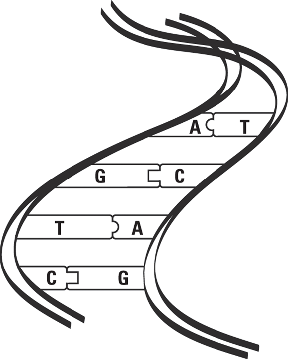
Illustration by Nan Owen
FIGURE 15-2: The DNA double helix.
The base pairing rule that C bonds only with G and A only with T is critical when DNA replicates or reproduces, ensuring that all new DNA strands are exact copies of the originals. The process actually is simple but elegant.
Mine, mine, all mine: Uniqueness and DNA
Each of us has approximately 3 billion base pairs (A-T and G-C pairings within the DNA molecules) in our DNA. That’s 6 billion bases in all. Because these bases can be put together in any order, the possible base sequences (series of bases linked in a linear fashion) for any given DNA strand are astronomical. That number, of course, is the basic reason that all humans are different, and it serves as the basis for DNA typing in the forensics lab.
With two notable exceptions — reproductive cells and red blood cells (RBCs) — all the cells in your body have 23 pairs of chromosomes within their respective nuclei. The red blood cells (RBCs) don’t have nuclei and thus have no chromosomes. Reproductive cells (eggs and sperm), on the other hand, contain only 23 unpaired chromosomes. These cells pair off with each other during fertilization, giving each new person a total of 46 chromosomes that contain genetic information, half from the father and half from the mother.
More than 8 trillion possible combinations arise from just two parents. (No wonder you don’t look like your sister.) The mother randomly donates one chromosome from each of her 23 pairs of chromosomes to each egg she produces. Which member of each pair she donates is independent of which member of every other pair she donates.
Of course, the same number of possibilities exists in the father. Because any sperm can combine with any of the mother’s eggs, the possibilities become huge.
Fingering Criminals Using DNA Fingerprints
Solving crime means finding out whodunit, so forensic scientists have long searched for ways to absolutely identify individuals from materials left at a crime scene.
The first discovery that provided positive proof was fingerprints, which are absolutely individual. No two people share the same prints, so fingerprinting became and remains an extremely powerful forensic tool. However, fingerprints aren’t found at every crime scene. Criminals have learned to wear gloves and to wipe their prints from any objects they touch.
Tracking down every bit of biological debris that gets left behind, however, is impossible for even the best criminals. DNA fingerprinting gives the criminalist an extremely accurate tool for using the tiniest bits of genetic material to identify individuals who were present at a crime scene.
Tracking down and preserving DNA
DNA is found in almost every cell in the human body. Skin, hair follicles, semen, saliva, and blood are common sources of crime-scene DNA. Hair doesn’t contain cells, but hair follicles do. Saliva doesn’t contain cells, but as it passes through the salivary ducts and washes around the mouth, it picks up cells from the ducts and the mouth. RBCs have no nuclei, so they contain no DNA. The DNA found when blood is tested comes from the white blood cells (WBCs). Using modern techniques, each type of fluid or tissue yields enough usable DNA for testing.
After it’s secured from a crime scene, DNA must be handled carefully to keep it from degrading. The best DNA samples are the ones that have been adequately dried and stored in protective containers. When drying isn’t feasible, wet samples need to be frozen until they’re analyzed.
Imagine trying to read a book in which all the sentences have been reduced to fragments or single words. War and Peace might be indistinguishable from Green Eggs and Ham. However, if the original books were merely torn into pages, you’d have little trouble distinguishing between the two. Similarly, DNA typing and matching depends upon the preservation of the sequence of the bases that make up the DNA. If the lab has only very short fragments or single bases to work with, much as a book reduced to a pile of single words, it can’t effectively type the DNA.
The bigger the DNA sample, the better, and yet usable DNA has been obtained from small and unlikely sources. Even a toothbrush, stamp, or bite wound can yield a usable saliva sample. A single drop of blood or a single hair follicle often is enough. In fact, with the newer techniques available to the DNA analyst, a single cell can supply enough DNA for testing.
In addition, modern technology makes possible the extraction of usable DNA from ancient tissues, even those taken from mummies that are thousands of years old. Scientists have extracted DNA from the bones and teeth of very old skeletal remains and, at times, from severely burned bodies (see the “Who’s yer granny and granddaddy? Mitochondria and Y-STR DNA” section later in this chapter).
Looking into the genome
The genome is the total DNA within the cell, or the millions of base pairs that make up the long polymers of DNA. Out of that massive number of base pairs, only about 5 percent directly carry out the work of life. These genes are encoded, meaning that they direct the synthesis (or manufacture) of proteins that the body needs for growth and function. The other 95 percent of the genome is non-encoded, which means it doesn’t directly code for the production of a protein, but it doesn’t simply lie around doing nothing, either. A portion of it regulates how genes function, and much of it is repetitive information whose purpose scientists haven’t yet been able to identify. These areas of non-encoded DNA are often called “junk DNA,” and it is these segments that are of primary interest to the forensic DNA analyst.
 All humans, and indeed all primates, share a large amount of the genome, meaning that much of your DNA is exactly like mine and everyone else’s. It’s also identical to that of the chimpanzees at your local zoo. Even so, that leaves plenty of unique combinations of DNA to give forensic investigators a keen method of finding out exactly who you are.
 In 1984, Alec Jeffreys and his associates at Leicester University discovered that each person’s DNA is actually unique. They found that certain areas of the long human DNA molecule exhibit polymorphism, a fancy word that means it can take many different forms. These variable areas are unique in everyone, and analyzing these areas allows scientists to make distinctions between one individual and the next. In 1985, shortly after discovering this polymorphism, Jeffreys developed a process for isolating and analyzing these areas of human DNA that he termed DNA fingerprinting. The process is also called DNA typing.
Polymorphisms important for forensics can be found in non-encoded, or junk DNA. These areas are highly variable in length and base sequence. The variability in length is called length polymorphism. It’s an important factor in forensic DNA typing because certain base sequences within the non-encoded DNA segments are constantly repeated. As a result, forensic investigators look for two types of sequences:
- Variable Number Tandem Repeats (VNTRs): The same base sequence repeats throughout a specific locus within the strand. These segments can be hundreds of base pairs long, repeating along the length of the DNA strand a variable number of times. VNTRs are rarely, if ever, used anymore as STRs and SNPs have supplanted their usefulness.
- Short Tandem Repeats (STRs): Much shorter than VNTRs — usually three to seven base pairs long — these sections also repeat throughout portions (loci) of the DNA chain. STRs repeat over segments of the DNA strand as long as 400 bases, which means that by using STRs, lab technicians can use even severely degraded samples for testing. Many more STRs are known than VNTRs, which gives forensic scientists many more repeats to analyze.
- Single Nucleotide Polymorphism (SNPs): SNPs are the most common DNA variations in humans, and there are literally millions of them scattered along our DNA strands. For example, a known location along the DNA strand might have a cytosine molecule in one person and an adenine or a thymine molecule in another person. A “match” is made if several of these are examined and a suspect’s SNPs match those of the crime scene sample. Because SNPs are much smaller than STRs, this technique can be useful in DNA samples too degraded for STR analysis.
The key in DNA typing is that the variability in the pattern of these repeats from person to person is broad, meaning that if technicians can isolate a certain locus of the DNA strand and determine the number of repeats of a given sequence in that area, they can compare it with another DNA strand to find out whether the pattern matches. In addition, research has determined how often a given number of repeats is found at a specific location in the DNA of the general population. Criminalists can use that information to calculate the probability that two DNA samples came from the same person. However, a match from a single locus is not very conclusive. But if several loci match, the probability quickly adds up.
Repeating yourself: How duplication identifies you
In terms of DNA, all of us repeat ourselves, but the specific ways in which we do it make each of us unique. When working to match DNA, investigators look at the repeats on particular loci of DNA. If my DNA were being compared to yours, for example, investigators would look at the same locus on each of our samples. They might find that you received 8 repeats of a particular STR from one parent and 14 repeats from another, and that I received 15 repeats of the same STR from one parent and 23 from another. Your DNA and mine would be very different.
But would your and my DNA be different from everyone else’s on Earth? You couldn’t tell by looking at only one locus. Other people also may have received 8 and 14 or 15 and 23 repeats for the same locus, but when you look at a dozen loci, the probability that two people received the exact number of repeats from each parent at all 12 loci is only one in several hundred trillion.
Looking at another example, if you were to analyze the STRs of a crime-scene sample at five different loci, you might find the following repeats:
|
Locus 1 |
12 and 9 |
|
Locus 2 |
6 and 14 |
|
Locus 3 |
23 and 16 |
|
Locus 4 |
5 and 18 |
|
Locus 5 |
8 and 19 |
Now, say that you already know that each of these STR repeat patterns occurs at these specific loci at respective rates of 1 percent, 3 percent, 8 percent, 1 percent, and 2 percent within the general population. That means 1 in 100 people share the same repeat pattern at Locus 1, 3 in 100 share this same repeat pattern at Locus 2, and so on. Therefore, if a suspect’s DNA shows the exact same repeat patterns at all five loci as the crime-scene sample, the probability that the DNA found at the scene came from someone other than the suspect is tiny. In fact, because the inheritance of the STR patterns at each locus is independent of any other locus, the percentages must be multiplied by each other to determine the probability of the DNA coming from someone other than the suspect, which in this case is a whopping 48 out of 10 billion, and that high degree of probability was found using only five loci. Imagine what those odds would be if the suspect’s DNA matched the crime-scene sample at 12 or more loci. Book him, Dano.
Understanding the DNA Fingerprinting Process
DNA evidence is incredibly reliable, but it requires some special treatment before it can be coaxed into revealing its secrets. Unlike fingerprint evidence, you can’t just “lift” a DNA sample and look at it in the right light to pull answers from it. Getting a DNA sample, and getting that sample into a form that allows investigators to analyze it and compare it with another sample, takes time and a lot of careful work.
The old method of DNA analysis was termed restriction fragment length polymorphism (RFLP) and required longer VNTRs. This method has been replaced by STR and SNP analyses in almost every jurisdiction. The reason is simply that RFLP required longer VNTRs (several hundred bases), while STRs are short (4 to 7 bases) and SNPs only a single base, or nucleotide. This means that the DNA to be analyzed can be more degraded and STR and SNP can still be useful as compared to using the old RFLP techniques.
Currently, the standard for DNA testing is STR analysis, coupled with the polymerase chain reaction (PCR). Using PCR, the DNA in even very small samples can be amplified (reproduced) at will. For this reason, the DNA from a single cell can be used to create a virtually unlimited quantity of DNA for testing.
SNP analysis offers an excellent complement to STR analysis, and current research suggests that it might even be able to distinguish identical twins — who by definition have the same DNA — from one another. The reason is that the continuing, lifelong replication of DNA within our cells as they synthesize new cells is subject to copy errors in single nucleotides. So over the years, the DNA of these twins develops variations that can often be exposed with SNP analysis.
This evolution in DNA analysis techniques was underlined by the famous Green River Killer case, as discussed in the sidebar “The Green River Killer.”
Getting even smaller with SNPs
An even newer technique, single nucleotide polymorphism (SNP), uses single nucleotides, meaning that the exact sequence, down to its actual base (nucleotide) sequence, is determined. This means that DNA analysis has evolved from using longer VNTRs to employing shorter STR repeats and now to determining the actual sequence of the bases in the analyzed sample. You can see this makes even more severely degraded samples useful to the DNA analyst.
SNP and similar determinations also make it possible for DNA analysts to distinguish one identical twin from the other. How is this possible? Don’t identical twins share the same DNA? Well, almost.
Fraternal twins come from different eggs and different sperm cells. That is, two eggs are fertilized by two different sperm cells at conception. These twins are as different as if born years apart. They are twins simply because they shared the mother’s womb prior to birth. Identical twins come from a single fertilized egg. In this case, when the fertilized egg undergoes its first division, the two daughter cells drift apart and then progress to become two distinct individuals. So these twins share the same DNA. Interestingly, even identical twins have different fingerprints, so if these are available, one can be distinguished from the other.
But even without fingerprints, the DNA analyst still has a trick or two to employ. In identical twins, as each of the two identical fertilized eggs produced by the first cellular division continues to multiply and ultimately grow into two different people, the replication of the DNA in each is not perfect. One twin will have a slightly different base sequence than the other. Though with FLRP and STR analysis they may appear identical, and thus indistinguishable by DNA fingerprinting, SNP analysis reveals these minute differences. Perpetrators can no longer blame their crime on their “evil identical twin.”
Making the match
Even though the graphs produced by RFLP and STR analysis look very different, they are actually the same. Each visually and numerically presents the DNA analysis in forms that are fairly easy to interpret. Matching the autoradiographic bands (RFLP) or the peaks (STR) produced from the analysis of a suspect’s DNA with that obtained from the crime scene can exonerate or implicate the suspect.
For example, say that a perpetrator shed blood at the scene of a homicide, and the crime lab found and collected it. The perpetrator’s blood constitutes an unknown sample because its origin isn’t known. When the suspect list includes two people, the lab takes a sample from each of them. The suspects’ samples are considered known samples because their origin is known. DNA fingerprinting compares the suspects’ DNA with the unknown crime-scene DNA.
In RFLP analysis, bands are created (Figure 15-3), whereas with STR, peaks are generated (Figure 15-4). Even a casual glance distinguishes a match from a non-match.
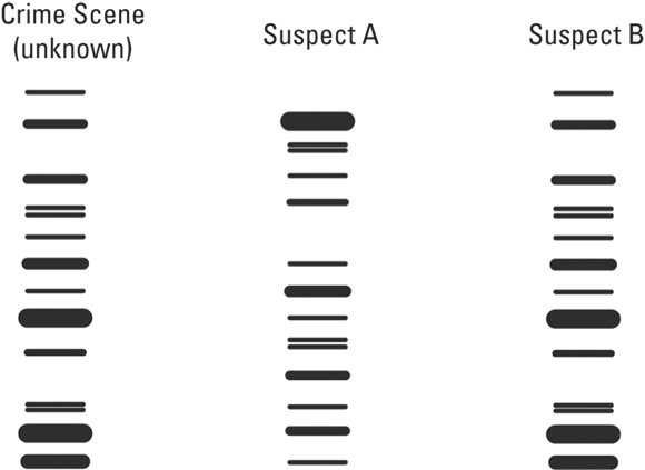
Illustration by Nan Owen
FIGURE 15-3: This autoradiograph result shows that the crime-scene DNA apparently came from Suspect B.
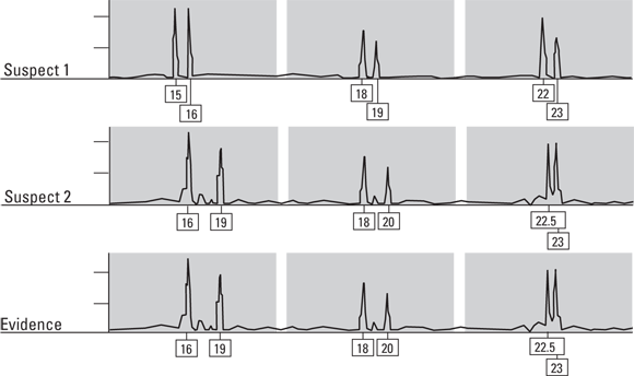
Illustration by Nan Owen
FIGURE 15-4: These STR peaks reveal that the evidence sample came from Suspect 2.
Shedding cells: Touch DNA
Did you know that you often leave behind your DNA simply by touching something? Fingerprints left at crime scenes consist of oils, dirt, and grime that has collected on the person’s finger pads. The print also contains shed skin cells, and these cells contain DNA. With PCR-STR, even a single cell deposited as part of a fingerprint can lead to a complete DNA profile. So, blood loss by the perpetrator isn’t necessary to grab a DNA profile; merely touching something at the crime scene will suffice.
These fingerprint materials can also be subjected to toxicological analysis (see Chapter 16 for the lowdown on toxicology) to reveal whether the person who deposited the print has used or handled substances such as cocaine or GHB.
Keeping it in the family: Familial DNA
As I state earlier, humans share much of the same DNA; the devil — and the crime lab techniques for analysis — lies in the minor variations. This is particularly true among family members. Brothers and sisters and parents and children share more DNA than do nonrelated individuals. Relatives’ DNA isn’t close enough for true DNA matching, but it’s close enough to suggest a connection.
Sometimes the evidence in an investigation strongly points to a particular suspect, but it’s not enough to secure an indictment and a successful prosecution. Often DNA from a suspect isn’t available, and there isn’t enough evidence to obtain a probable cause warrant for investigators to demand a sample. In such situations, investigators may turn to familial DNA by testing the DNA of close family members. If the DNA profile of these individuals is close to the crime-scene sample, this may be enough to get that needed warrant and force the suspect to submit a sample.
Using DNA to Determine Lineage
You have more than your father’s ears and your grandmother’s hooked nose — you have the DNA that they passed down to you. Because DNA is so individual and because it’s passed down from one generation to the next, it’s an extremely powerful tool for determining an individual’s ancestral background. Whether it’s proving or disproving fatherhood or tracking distant ancestors, DNA testing is a reliable tool that provides compelling evidence.
Who’s yer daddy? Paternity testing
ABO blood typing can be used to exclude paternity, but it can’t absolutely state that a certain man is the father of a certain child (see Chapter 14). DNA testing, on the other hand, can confirm paternity. To determine paternity using DNA, scientists make profiles of the mother’s DNA and the child’s DNA. These profiles then are compared to the profile of the suspected father. The child’s DNA pattern must be a combination of the DNA profiles of the mother and father. The child’s DNA profile can’t include a band, or peak, that neither parent supplies; however, the child’s profile doesn’t necessarily have every band that each parent possesses. Remember, each parent only gives half of the offspring’s DNA. But, because all of the child’s DNA must come from its parents, the child can’t have DNA that neither of them has.
Through the union of egg and sperm, each parent donates half of her or his DNA to the new child. Neither, however, donates all of his or her DNA. Chromosomes are arranged in 23 pairs, and each parent donates one chromosome of each pair to the child. Thus, the child is a combination of DNA donated by each parent. Parents may have DNA that the child doesn’t have (DNA on the chromosome not donated via the egg or sperm), but the child absolutely can’t have DNA that wasn’t donated by at least one of the parents. So when the child’s DNA is fragmented and separated into bands or peaks, each of the resulting bands or peaks must also be found in the DNA profile of one or the other parent.
In paternity testing, if the child possesses a DNA fragment that isn’t present in either the mother or the suspected father, then the man is not the child’s father. This fragment must have come from someone else (the true father), and paternity for the suspect father is excluded. (See Figure 15-5.)
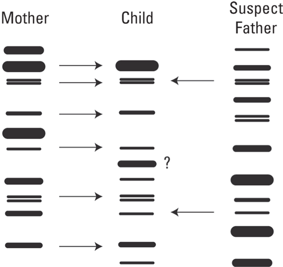
Illustration by Nan Owen
FIGURE 15-5: This child has a band that isn’t found in the DNA profile of either the mother or the suspect father, who can’t be the actual father.
Who’s yer granny and granddaddy? Mitochondrial and Y-STR DNA
Mitochondrial DNA adds an extremely useful tool to the forensic toolbox. It helps in identifying perpetrators and human remains and for determining ancestry.
 The DNA used in standard DNA testing is nuclear DNA. It can be extracted from any nucleated cell. But cells also contain non-nuclear DNA. This DNA is found within the mitochondria. Mitochondria are small organelles that reside within the cytoplasm of the cell and serve as the cell’s energy production center. A small amount of DNA is in the mitochondria, but each cell has many mitochondria organelles. Mitochondrial DNA is important for several reasons, including the following:
- Passes from generation to generation by the maternal lineage
- Mutates rarely
- Is found in places where nuclear DNA doesn’t exist
- Is exceptionally hardy
Your mitochondrial DNA (mtDNA) is inherited unchanged from your mother and only from your mother. She received hers from her mother, and her mother from her mother, and so on.
At fertilization, the mother’s egg supplies the cell and half the DNA, but the sperm supplies only half the DNA. The sperm cell breaks down and disappears after passing its genetic material into the nucleus of the egg cell. Thus, all the cell components of the developing zygote (fertilized egg) come from the mother, including the mitochondria. As the cell divides and multiplies, these mitochondria are copied and passed on, generation after generation, and that means all the cells of the body contain identical mtDNA.
The rare mtDNA mutation is thought to occur approximately once every 6,500 years. Thus, mtDNA is extremely stable, and that means that your mtDNA is virtually identical to that of your mother, your great-great grandmother, and your maternal ancestors from a thousand years ago. Thus, you can accurately trace your maternal lineage across many generations.
Y-STR DNA is similar to mDNA except that it is located on the male Y chromosome and is passed down along the paternal ancestry line. This means that a male would share his Y-STR DNA with his father, brothers, paternal grandfather, paternal uncles, and paternal nephews if born from a paternal uncle rather than a paternal aunt.
Because mtDNA is hardy, scientists can often extract it from the bones and teeth of very old skeletons and thus use it to identify and determine the ancestry of skeletal remains. Mitochondrial DNA also is found in some tissues where nuclear DNA can’t be found. For example, hair predominantly is composed of dead cellular debris. The only living part of hair is the follicle. The cells of the follicles contain nuclear DNA, which can be used for DNA profiling and matching. In other words, hair that’s been yanked out or shed with its follicular bulb attached can provide nuclear DNA.
But, what if the hair has been cut, and no bulb is attached? All is not lost.
As hair grows, the cells of the bulb multiply, undergo change, and become incorporated into the growing hair. Part of this transformation is the loss of the nucleus from each cell. Thus, although hair has no nuclear DNA, the dead cellular debris that is incorporated into the hair shaft does contain mtDNA that can be extracted and used for identifying the person who shed the hair.
If a hair is found at a crime scene and it matches the suspect’s hair in every physical detail, investigators may reasonably suspect that he shed the hair while at the scene. The problem is that such “proof” is shaky at best. But if the mtDNA from the crime-scene hair matches that of the suspect, the evidence becomes more powerful because the crime-scene hair must have come from the suspect or at least someone who shared the suspect’s maternal ancestry.
Indexing DNA: The CODIS system
The FBI’s CODIS Unit oversees the Combined DNA Index System (CODIS) and the National DNA Index System (NDIS), which are databases of DNA fingerprints taken from felons and from biological fluids obtained from crime scenes, such as assaults, homicides, and rapes. Forensic scientists hope the database becomes the DNA equivalent of the Automated Fingerprint Identification System (AFIS).
In the same way that a fingerprint can be entered into AFIS and matched against a database of fingerprints from across the country, DNA profiles can be compared through CODIS. Unfortunately, the system isn’t yet used in every state and jurisdiction. The FBI is in the process of expanding it into a truly nationwide system.
With CODIS, criminalists are able to compare a crime-scene DNA sample with profiles of known felons and DNA obtained from other crime scenes to establish matches. The DNA in question may match DNA obtained from another crime scene and link the crimes. As a result, evidence from crime scenes can be considered together, which may lead to the identity of a perpetrator. As witnessed by the seemingly constant stream of cold cases solved by DNA matching, CODIS already has enjoyed many successes.