Jianjun Cheng and Suzie Hwang Pun
Therapeutic drugs are usually delivered in bolus doses, either orally or by injection. These administrations result in initial blood concentrations that are higher than required levels, followed by a decrease to subtherapeutic levels due to drug degradation and excretion. Therefore, drugs must be given frequently to maintain therapeutic drug concentrations (Figure 16-1). In the 1970s, researchers introduced the concept of controlled drug delivery by using carriers to release drugs in a sustained manner. In the ideal case, drugs are gradually released from a depot so that the drug concentration is maintained at an effective level over a long period. An example of one such success is Gliadel, the first FDA-approved biopolymer drug-delivery system for treatment of brain cancer. Gliadel wafers are implanted into the brain after tumor resection. There, the wafers locally release carmustine (a chemotherapeutic drug) for several months.

Many other promising drugs never make it to clinical trials because of inherent pharmacological drawbacks. Low-molecular-weight drugs, such as most chemotherapy drugs, are usually insoluble and highly toxic. Protein and nucleic acid drugs usually have poor stability in physiological conditions. It is therefore essential for these drugs to be protected en route to their target disease sites in the body. Drug-delivery systems may rescue potential drug candidates by increasing solubility and stability.
Drug-delivery technologies are developed to improve the safety and efficacy of drugs, to ensure better patient compliance, and to improve the shelf life and stability of therapeutic products. Controlled drug release involves the combination of a biocompatible material or device with a drug to be delivered in a way that the drug can be delivered to and released at diseased sites in a designed manner.
The major routes of drug administration are oral, inhalation, injection, and transdermal delivery. The most well-known route is oral drug delivery, which accounted for about 50 percent of the market as of 2003. The other routes of administration—inhalation, transdermal, injection and implantation, and nasal delivery—account for the remaining market share at 19 percent, 12 percent, 10 percent, and 7 percent, respectively. In the past 30 years, the field of drug delivery has been undergoing rapid development and has attracted attention from both academia and pharmaceutical industries. According to a recent report, the U.S. market alone for drug delivery is estimated at $43.7 billion in 2003 and is expected to grow more than 11 percent annually in the next five years.[1]
Nanotechnology, a multidisciplinary area of science involved in the miniaturization and use of materials or devices on the nanometer scale, has been undergoing explosive growth and has become a principal research interest. In the past ten years, nanotechnology has already demonstrated great impact in almost every frontier area of drug delivery by extending and improving traditional delivery techniques.
One of the earliest applications of nanotechnology in drug delivery was conceived in the 1970s, when nanoparticles were designed as carriers of anti-cancer drugs. Since then, many nanoparticle systems have been developed for use in drug delivery. Biopolymers have been intensively studied for application in nanoparticulate drug delivery.
Delivery vehicles involved with polymeric systems include polymerdrug conjugates, polymeric micelles, polymeric nanospheres and nanocapsules, and polyplexes. Inorganic and metallic materials have also been used in preparation of nanoparticles for drug delivery. Recently nano- and microfabrication technologies have been applied for drug delivery, resulting in novel devices such as biochips and microneedles. With the explosive growth in the development of new methods of nanofabrication, numerous emerging nanosystems will inevitably change the field of drug delivery in the coming decades.
Nanoparticle-based delivery vehicles improve drug efficacy by modulating drug pharmocokinetics and biodistribution. Small-molecule drugs are rapidly eliminated from the circulation by the kidneys. Injectable nanoparticledelivery vehicles, typically ranging from 5nm to 200nm in size, substantially increase circulation (particles >5nm avoid kidney clearance) while minimizing removal by cells that police the blood for foreign particles (macrophages have less propensity for particles <200nm in size). Oral delivery is currently the most preferred method of drug administration because of its cost effectiveness and ease of use.
The market for oral drug-delivery systems has been growing at a rate of 8.6 percent per year since 2000. A major area of research in oral delivery is in delivery materials for protein drugs. Because particle permeability across the intestinal wall is inversely proportional to size, nanoparticles used for oral delivery offer obvious advantages. The interest in nanoparticle-based drug delivery for other administration routes is also growing. The following sections focus on polymer, lipid, and inorganic or metallic nanoparticles that are <500nm in size.
Polymer-drug conjugates (5–20nm) represent the smallest nanoparticulate delivery vehicles. The polymers used for such purposes are usually highly water-soluble and include synthetic polymers (for example, poly(ethylene glycol) (PEG)) and natural polymers (such as dextran). When hydrophobic small molecules are attached to these polymers, their solubilities in water can be substantially improved. For example, a cyclodextrin-based polymer developed at Insert Therapeutics increases the solubility of camptothecin, an insoluble chemotherapy drug, by three orders of magnitude.
Small molecules or proteins conjugated to these polymer delivery vehicles can achieve extended retention in circulation because of reduced kidney clearance. PEG-L-asparaginase (ONCASPAR; Enzon), an FDA-approved PEGylated protein drug as a treatment for acute lymphoblastic leukemia, can be administered every two weeks, instead of the two to three times per week required for the non-PEGylated enzyme. Other PEGylated systems approved by the FDA include PEG–adenosine deaminase (ADAGEN; Enzon) as a treatment for X-linked severe combined immunogenicity syndrome and PEGinterferon (PEGASYS; Roche and PEG–INTRON; Schering-Plough) as treatments for hepatitis C.[2] Many polymer-small molecule and polymer-protein conjugates are currently in clinical trials. A promising nanoparticle drug-delivery system—a proprietary, albumin-bound paclitaxel conjugate codeveloped by American Pharmaceutical Partners and American BioScience—has shown excellent antitumor efficacy in Phase III clinical trial for the treatment of metastatic breast cancer.
One group of polymers that has attracted enthusiasm recently are dendrimers (Figure 16-2). They are monodispersed, symmetric, globular-shaped macromolecules comprising a series of branches around an inner core. Dendrimers are potential nanometer-sized systems for drug delivery, and their sizes can be controlled simply by adjusting the generation of dendritic branches.

Figure 16-2. Schematic drawing of dendrimer for application in drug delivery and targeting. (Reprinted with permission from http://www.drugdeliverytech.com/cgi-bin/articles.cgi?idArticle=153, Thiagarajan Sakthivel, Ph.D., and Alexander T. Florence, Ph.D., D.Sc., “Dendrimers & Dendrons: Facets of Pharmaceutical Nanotechnology.”)
Amphiphilic block copolymers—polymers that contain both hydrophilic and hydrophobic regions—tend to self-assemble in aqueous solution into spherical structures called micelles. Polymeric micelles typically have a hydrophilic corona and a hydrophobic shell. When used as drug-delivery agents, polymeric micelles are most commonly formulated to include hydrophobic drugs (for example, doxorubicin, cisplatin, amphotericin B) in the core, leaving the outer hydrophilic layer to form a stable dispersion in aqueous media.
The stable structure of the polymeric micelles prevents rapid dissociation (release of drug) in vivo. Polymer micelles typically range from 60 to 100nm, with fairly narrow size distributions. The micellar corona can be further modified with targeting moieties (for example, antibodies) to deliver drugs to desired sites. Development of micellar drug-delivery vehicles is in an early stage, with most formulations still in preclinical studies.[3]
Nanoparticles are solid, small colloidal particles made of polymers having diameters from 50nm to several hundred nanometers. Depending on the method of preparation, two types of drug-containing nanoparticles exist: nanospheres (a matrix system in which drugs are uniformly distributed) or nanocapsules (a reservoir system in which drugs are confined to the core of the particles and are surrounded by polymer membranes). Many of these systems are made of biodegradable polymers, such as poly(ortho ester) (Figure 16-3). Poly(ortho ester) nanoparticles release drugs with tunable release rates, depending on solution pH. Drugs encapsulated in nanoparticles have increased stability against enzymatic and chemical degradation, an important advantage for unstable drugs such as proteins and nucleic acids.[4]

Figure 16-3. Scanning electron microscopy image of poly(ortho ester) nano- and microspheres. Scale bars: 5mm. (Reprinted with permission from Chun Wang, Qing Ge, David Ting, David Nguyen, Hui-Rong Shen, Jianzhu Chen, Herman N. Eisen, Jorge Heller, Robert Langer, and David Putnam, “Molecularly engineered poly(ortho ester) microspheres for enhanced delivery of DNA vaccines,” Nature Materials 3 (2004): 190–196.)
Nanoparticles have been tested for the delivery of all types of drugs (small molecules, proteins, and nucleic acids) in almost all types of administration routes (such as inhalation, oral, and injection). Although many of these approaches are still in an early stage of their development, some of them have already shown great potential. An example is Dr. Edith Mathiowitz’s (Brown University) poly(fumaric-co-sebacic) anhydride nanoparticles for the oral delivery of insulin, a promising way to achieve oral protein delivery.[5]
In nonviral gene therapy, plasmid DNA is introduced into cells to express therapeutic proteins, whereas in oligonucleotide therapy, oligonucleotides (such as ribozymes and DNAzymes) and small, interfering RNA (siRNA) are used to suppress disease-associated expression. However, the cell membrane is a natural barrier for these genetic materials. For therapeutic nucleic acids to be successfully delivered into the cell, they must be complexed with materials that facilitate cellular uptake.
Polyplexes, a group of nanoparticulates formed by charge interaction between positively charged polymers and negatively charged nucleic acids, are developed for such purposes. Polyplexes range in size from 40nm to 200nm (Figure 16-4). RNA interference (RNAi) is an emerging and promising approach for oligonucleotide therapy, and there is currently active research in developing materials for siRNA delivery.
![Cyclodextrin polycation-based polyplexes developed by Davis and coworkers (California Institute of Technology).[6] These materials are being investigated for gene therapy applications at Insert Therapeutics. Bar is 100nm. (Reprinted with permission from S. J. Hwang, N. C. Bellocq, and M. E. Davis, “Effects of Structure of beta-cyclodextrin-containing Polymers on Gene Delivery,” Bioconjugate Chemistry 12(2) (2001): 280–290.)](http://imgdetail.ebookreading.net/math_science_engineering/11/0131927566/0131927566__nanotechnology-science-innovation__0131927566__graphics__16fig04.jpg)
Figure 16-4. Cyclodextrin polycation-based polyplexes developed by Davis and coworkers (California Institute of Technology).[6] These materials are being investigated for gene therapy applications at Insert Therapeutics. Bar is 100nm. (Reprinted with permission from S. J. Hwang, N. C. Bellocq, and M. E. Davis, “Effects of Structure of beta-cyclodextrin-containing Polymers on Gene Delivery,” Bioconjugate Chemistry 12(2) (2001): 280–290.)
Liposomes—nano-sized particles (25 to several hundred nanometers) made from phospholipids and cholesterols—are sometimes referred to as “fat bubbles.” Liposomes consist of bilayers of lipids that can encapsulate drugs. Their properties for use as delivery vehicles are closely associated with lipid composition, liposomal size, and fabrication methods. For example, saturated phospholipids with long hydrophobic chains usually form a rigid, impermeable bilayer structure, whereas the unsaturated phosphatidylcholine-based lipid layers are much more permeable and less stable.
Liposomal drug delivery has achieved great success in the past decade. Several liposome-based drug-delivery systems have been approved for clinical use. AmBisome (lipid-based delivery of amphotericin B, Fujisawa Healthcare, Inc., and Gilead Science) was approved for the treatment of cryptococcal meningitis in HIV-infected patients. The sales of AmBisome were nearly $200 million in 2003 (an increase of 7 percent from 2002). Doxil (Alza) was approved in 1999 for the treatment of refractory ovarian cancer and is the first and only liposomal cytotoxic agent approved to treat a solid tumor. The sales of Doxil reached $80 million in 2000.
Delivery of drugs using new inorganic and metallic nano-sized vectors are still in the proof-of-concept of stage. One unique approach originates from C-60, a soccer ball–shaped fullerene.[7] Another interesting approach is to use magnetic nanoparticles to carry chemotherapeutic drugs to cancer sites directed by an external magnetic field.
Very recently, other metal nanoparticles have been investigated as therapeutics and drug-delivery systems. An example from Dr. Naomi Halas’s research group (Rice University) is the nanoshell, a new type of nanoparticle composed of a dielectric silica core coated with an ultrathin gold layer.[8] Once the nanoshells penetrate tumor tissues, they can be activated for thermal therapy by taking advantage of their ability to convert absorbed energy from the near-infrared region to heat.
Implantable drug delivery (IDD) devices offer more uniform drug release rates, lower required doses, and localized delivery. These devices are generally composed of the drug of interest distributed in a polymer matrix. Examples of marketed implantable drug-delivery formulations include Norplant for birth control (Wyeth Laboratories), Gliadel for localized delivery of a chemotherapeutic agent to the brain (Guilford Pharmaceuticals), and Viadur for slow release of hormones for prostate cancer treatment (Bayer). The sales of Gliadel in 2003 were nearly $20 million (an increase of 32 percent from 2002), and annual Viadur sales are projected to reach $150 million.
Based on these trends, the demand for implantable drug-delivery systems is expected to exceed $2 billion by 2012. There is still room for improvement in IDD technology. Two emerging nanotechnologies with applications in implantable drug delivery are nanoporous membranes and biochips.
Nanoporous membranes are microfabricated with well-defined pores (diameters in the tens of nanometers). The membranes can be used to deliver small-molecule, peptide, or protein drugs (Figure 16-5). One application under investigation involves encapsulation of pancreatic islet cells for insulin delivery. The reproducible and uniform pore size precisely controls the material exchange across nanoporous membranes: Nutrients for the cells and secreted insulin can pass through the pores, but proteins and cells from the immune system that may attack the implanted islet cells are restricted from entering the biocapsules due to their size.
![Nanoporous membranes developed by Desai and colleagues have nanometer-sized pores for controlled material exchange.[9] (Reprinted with permission from S. L. Tao and T. A. Desai, “Microfabricated drug delivery systems: From particles to pores,” Advanced Drug Delivery Reviews 55 (2003): 315–328.)](http://imgdetail.ebookreading.net/math_science_engineering/11/0131927566/0131927566__nanotechnology-science-innovation__0131927566__graphics__16fig05.jpg)
Figure 16-5. Nanoporous membranes developed by Desai and colleagues have nanometer-sized pores for controlled material exchange.[9] (Reprinted with permission from S. L. Tao and T. A. Desai, “Microfabricated drug delivery systems: From particles to pores,” Advanced Drug Delivery Reviews 55 (2003): 315–328.)
Drug release from IDD devices is not constant, and “burst” effects are still observed. In addition, drug release cannot be controlled after implantation of IDD devices. Biochips have been developed to precisely control the amount of drug released. Biochips are usually fabricated using silicon and contain a number of mini-wells with precisely controlled sizes, each capable of holding a few hundred nanoliters. These mini-wells are loaded with drugs and are covered with caps of thin metal foils (usually gold) that are connected to the wires on the face of the chips (Figure 16-6).[10] When electrical signal is applied, the current dissolves the metal covers and releases the drug. Biochips can be implanted beneath the skin or into more specific areas such as the spinal cord or brain. The electronics package outside the chips receives a radiofrequency instruction through a built-in antenna to order a microprocessor to control the melting of metal foils. MicroCHIPS, Inc., is one of the key companies developing this technology. Preclinical studies in animals using biochips developed by MicroCHIPS, Inc., have shown good biocompatibility without significant side effects. Once successfully developed, the biochip-based drug-delivery technology will allow for precisely controlled drug administration to patients.

Figure 16-6. A biochip for controlled release developed by Robert Langer and coworkers. (a) Schematic drawing of biochip; (b) a single reservoir. (Reprinted with permission from J. T. Santini, M. J. Cima, and R. Langer, “A controlled-release microchip,” Nature 397, no. 6717 (Jan. 28, 1999): 335–338.)
The first transdermal patch technology for drug delivery was approved by the U.S. FDA in 1979. In the past quarter-century, the U.S. market for transdermal patches has grown to more than $3 billion per year.[11] Transdermal drug delivery is an attractive, noninvasive approach to drug administration and is likely to improve the bioavailability of drugs by avoiding first-pass liver metabolism and by maintaining more uniform drug plasma levels than bolus injections.
Despite these advantages, successful application of transdermal patches has been largely limited to a handful of drugs, including nicotine (for smoking cessation), scopolamine (for motion sickness), and hormone replacement. The human skin acts as a protective barrier to prevent entry of most molecules. A lipid-rich layer in the skin repels water-soluble molecules, and large molecules are also largely excluded because of their size. Thus, most major families of drugs (hydrophilic small molecules, peptides, proteins, and nucleic acids) have not been successfully delivered by traditional transdermal patch technology.
The permeability of the skin to hydrophilic or macromolecule drugs can be increased by physically creating microscopic holes in the skin using arrays of microneedles. Ideally, the microneedles would create pores to facilitate drug entry but would remain small enough to be painless. Although technology was conceived several decades ago, the technology to prepare these systems was not available. Microfabrication technology, developed by the microelectronics industry, recently has been used to prepare these structures.
Prausnitz and colleagues first demonstrated this concept in 1998 by preparing silicon-based, solid microneedles (Figure 16-7). Since then, many variations of these structures have been prepared and tested with diverse drug families. Materials used to synthesize microneedles include glass, polymers, metal, and silicon. The microneedles have been successfully applied to deliver small molecules, oligonucleotides, plasmids, and proteins through the skin, increasing the permeability of these molecules by several orders of magnitude. Application approaches include using solid microneedles to pierce holes in the skin, followed by application of a traditional patch; coating solid microneedles with drugs that are slowly released from the needles after skin penetration; and flowing drug solutions through hollow microneedles.
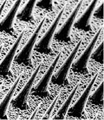
Figure 16-7. Solid silicon microneedles with heights ~150nm can be applied painlessly to enhance transdermal drug delivery. (Reprinted with permission from M. R. Prausnitz, “Microneedles for transdermal drug delivery,” Advanced Drug Delivery Reviews 56, no. 5 (2004): 581–587.)
A pilot human trial revealed that the microneedle arrays are indeed applied painlessly.[12] Transdermal microneedle delivery was quickly adopted by the pharmaceutical and drug-delivery industries; several companies are currently developing this technology for transdermal drug administration. As this technology matures, it has the potential to quickly surpass the market currently occupied by traditional patch formulations.
Nanotechnology has played a large role in advancing the drug-delivery field by enhancing existing areas of small-molecule and protein delivery and by opening doors for delivery of new families of nucleic acid-based drugs. The ability to control the properties of nanoscale materials will continue to impact the pharmaceutical field by providing new technologies for improved drug delivery. We are optimistic that these developments will lead to delivery vehicles with high target specificity and with the ability to precisely control drug release.