Chapter 3
MANUAL MATERIALS HANDLING
Robert B. Dick, Stephen D. Hudock, Ming-Lun Lu, Thomas R. Waters†, and Vern Putz-Anderson*
Manual handling is defined by the International Organization for Standardization as any activity requiring the use of human force to lift, lower, carry or otherwise move, or restrain an object, including humans and animals.1 Manual materials handling (MMH) excludes animate items as objects in such activities. The main risk factors associated with the development of injuries with MMH tasks are forceful exertions, awkward postures, repetitive motions, pressure points, and static postures.2 Many different types of jobs or occupations require MMH activities including moving household goods, warehouse handling, truck unloading, inventory restocking, production line loading and unloading, baggage handling, container loading and emptying, transferring material goods, and delivering packages.
Nongovernmental researchers annually examine US Bureau of Labor Statistics (BLS) injury data to ascertain which workplace events caused an employee to miss six or more days of work. Those workplace events are then ranked by total workers’ compensation costs. For the year 2012, the most recent information available, the leading cause of disabling injury was overexertion involving outside sources. This event category includes injuries resulting from lifting, pushing, pulling, holding, carrying, or throwing, all primarily MMH activities. These events cost US businesses $15.1 billion in direct costs and accounted for over 25% of the overall national disabling injury burden.3
In 2011 approximately 3 million workers in private industry and 8 21 000 in state and local government experienced a nonfatal occupational injury or illness with an estimated cost to the US economy of $200 billion annually.4 High-risk occupations, which are defined as one where the days-away-from-work (DAFW) rate is at least twice the DAFW rate of 113.3 cases of injury and illnesses per 10 000 full-time employees (FTEs), included two occupations with more than a million workers where MMH activities are involved. These were drivers (sales and trucks) and hand laborers (freight, stock, and material movers).4 The estimated number of drivers was 2 721 000 and the DAFW rate was 329.4/10 000 FTE, and for hand laborers the number employed was 1 616 000 and the DAFW rate was 440.3/10 000 FTE.4
Musculoskeletal disorders (MSDs), as defined by the BLS, include soft tissue injuries to the trunk and upper and lower extremities and occurred at a rate of 35.8 days-away-from-work cases per 10 000 FTE in 2013 (including private and state and local government).5 The MSD rate for transportation and warehousing was 80.3 cases per 10 000 full-time workers which was more than twice the MSD rate for all private industry sectors. MSDs accounted for 33% of all reported illness and injury cases in 2013. Nursing assistants and laborers and freight, stock, and material movers showed the greatest number of MSD cases in 2013.
This chapter provides an overview of the hazards associated with MMH. It will focus on muscular strains and sprains, primarily in the torso or extremities. The prevention of these injuries requires sufficient knowledge to both identify workplace hazards and implement changes in the job or process that will reduce or eliminate these hazards.
OCCUPATIONAL SETTING
MMH poses a risk of injury to many workers; injury is more likely to occur when workers perform tasks that exceed their physical capacities. In addition, the physical capacities of individual workers vary substantially. Because MMH hazards are present in many industrial and service operations, workers in a wide variety of industries are potentially at risk.
The BLS reports that in 2013 there were 1.6 million DAFW cases in private industry and state and local government.5 The overall incidence rate of nonfatal occupational injury and illness cases requiring DAFW to recuperate was 109.4 cases per 10 000 FTE. When the BLS data is broken down by body part, the incidence rate for trunk injuries is 26.4 per 10 000 FTE (20.0 for back). The rate for lower extremity (e.g., knee, ankle, foot) is 24.8 and upper extremity (e.g., shoulder, arm, wrist, hand) is 32.5.5
A recent report by the National Safety Council indicated that the total number of nonfatal private industry occupational injuries and illnesses involving DAFW was 905 690 cases in 2012.6 Overexertion was reported for 3 31 130 cases, and lifting and lowering activities accounted for 106 210 of these cases. On a percentage basis, the parts of the body affected that related to MMH activities were trunk (including the back) (20.4%), upper extremities (shoulder, arm, wrist, hand) (23.9%), lower extremities (knee, ankle, foot, or toe) (18.1%), and neck (1.3%). Transportation and warehousing, the industry sector where MMH activities prevail, had 89 260 nonfatal DAFW cases in 2012. On a percentage basis, the parts of the body affected that related to MMH activities were trunk (including the back) (21.7%), upper extremities (shoulder, arm, wrist, hand) (21.8%), lower extremities (knee, ankle, foot, or toe) (19.5%), and neck (2.1%). The average total workers’ compensation incurred costs per claim for parts of the body associated with MMH activities for 2011–2012 reported by the National Safety Council were as follows: (i) arm/shoulders, $42 742; (ii) lower back, $38 492; (iii) upper back, $34 297; (iv) multiple trunk/abdomen, $22 361; and (v) hand/fingers/wrist, $21 726.6 The actual percentage of workers employed in jobs that require MMH tasks is difficult to determine, but three recent surveys of the American workforce indicate that a large percentage of workers are in jobs that require heavy lifting and/or hand movements, which are two indicators of MMH. The three surveys were conducted in 2002, 2006, and 2010 and roughly indicate that 48% of respondents had jobs that required these activities.7–9
NORMAL ANATOMY AND PHYSIOLOGY OF THE SPINE
The spine is a complex structure made up of bony, muscular, and ligamentous components. The spine can be divided into two major subsystems—the anterior and posterior spine. The anterior spine is mainly composed of the large bony vertebral bodies. These vertebral bodies rest atop one another and are separated by the cartilaginous intervertebral disks, which act as “shock absorbers.” The vertebral bodies and disks are held together by two sets of ligaments. The posterior spine is made up of the additional bony structures of the vertebral peduncles and laminae, which together form the spinal canal. The facet joints, which join two adjacent vertebrae, and the lateral and posterior spinous processes also form part of the posterior spine. The spinous processes are the attachment points for muscles that move and support the spine.
The spine is dependent on both bony and nonbony support for stability. Bony support is provided by the intervertebral disks and the facet joints. Nonbony support comes from the ligaments and the attached musculature. As the bony structures and ligaments do not have enough strength to resist the forces generated during movement and lifting, the spine is dependent on the muscles of the back, abdomen, hip, and pelvis for stability. This principle explains why muscular fatigue is so important in the pathophysiology of back injury. The parts of the spine with the greatest degrees of movement are at highest risk. Because the thoracic and sacral vertebrae are fixed in place by the ribs and the pelvis, the lumbar vertebrae are the most common sites of injury. An excellent compilation of the anatomy and clinical conditions involving the lumbar spine is available in another publication.10
PATHOPHYSIOLOGY OF INJURY AND RISK FACTORS
The interpretation of the research linking work-related MSDs and MMH is problematic because of the high prevalence of certain disorders in the general population, such as low back pain (LBP), which have a frequent association with nonoccupational factors. In addition, the relationship is further obscured by the wide range of disorders, the nonspecific nature of the condition, and the general lack of objective data relating different risk factors to overexertion injury. In 1997, the National Institute for Occupational Safety and Health (NIOSH) published an extensive review of the epidemiologic literature that assessed the strength of the association between specific work factors and certain upper extremity and lower back MSDs. The NIOSH researchers identified more than 2000 studies, examined more than 600 epidemiologic studies, and published a comprehensive review of the epidemiologic studies of back and upper extremity MSDs and occupational exposures.11
A 2001 report by the National Research Council’s Institute of Medicine summarized a review of the literature on work-related risk factors for low back pain by stating “there is a clear relationship between low back disorders and physical load imposed by manual material handling, frequent bending and twisting, physically heavy work and whole body vibration.”12 A systematic review conducted by da Costa and Vieira13 examined 1761 studies published since the NIOSH11 review on work-related musculoskeletal disorders and concluded that there was reasonable evidence for causal relationship with heavy physical work and MSDs. The most frequently reported risk factors for MSDs were excessive repetition, awkward postures, and heavy lifting, which are activities involved in MMH.13
It is generally recognized that musculoskeletal injuries are a function of a complex set of variables, including aspects of job design, work environment, and personal factors. Moreover, work-related MSDs may result from direct trauma, a single exertion (overexertion), or multiple exertions (repetitive trauma); typically, it is difficult to determine the specific nature of the causal mechanism. A variety of manual handling activities increase a worker’s risk of developing an MSD, including jobs that involve a significant amount of manual lifting, pushing, pulling, or carrying and jobs requiring awkward postures, prolonged standing or sitting, or exposure to cyclic loading (whole-body vibration). In addition to these frequent patterns of usage, a variety of personal and environmental factors may affect the risk of developing an MSD. Risk factors increase the probability of occurrence of a disease or disorder, though they are not necessarily causal factors. For the problem of low back injuries involving MMH, four categories of risk factors have been identified by epidemiologic studies—personal, environmental, job related, and psychosocial/organizational:
- Personal risk factors are conditions or characteristics of the worker that affect the probability that an overexertion injury may occur. Personal risk factors include attributes such as age, gender, level of physical conditioning, strength, and medical history.
- Environmental risk factors are conditions or characteristics of the external surroundings that affect the probability that an overexertion injury may occur. Environmental risk factors include attributes such as temperature, lighting, noise, vibration, and friction at the floor.
- Job-related risk factors are conditions or characteristics of the MMH job that affect the probability that an overexertion injury may occur. Job-related risk factors include attributes such as the weight of the load being moved, the location of the load relative to the worker when it is being moved, the size and shape of the object moved, frequency of handling, and the grip forces required to move an object. These factors should be identified because they may be very important for preventing the magnitude of physical hazards to the worker.
- Psychosocial/organizational factors are factors related to the social or organizational environment such as occupational stress, job satisfaction, monotonous work, social support at work, perceived work demands, etc. The mechanisms for how these factors might increase the risk of MSDs is not fully understood, but research shows strong associations with increased physiological and biomechanical responses with MMH activities.7–9
A tabular presentation of risk factors is provided in Table 3.1.
TABLE 3.1 Risk factors associated with manual material handling injuries.
| Personal factors | Environmental factors | Job-related factors | Psychosocial factors |
| Gender | Humidity | Location of load relative to the worker | Occupational stress |
| Anthropometry (height and weight) | Light | Distance object is moved | Job satisfaction |
| Physical fitness and training | Noise | Frequency and duration of handling activity | Monotonous work |
| Lumbar mobility | Vibration | Bending and twisting | Social support at work |
| Strength | Foot traction | Weight of object or force required to move object | High perceived work demands |
| Medical history | Coupling/grip strength required | Job control | |
| Years of employment | Stability of the load | Time to complete work tasks | |
| Smoking | |||
| Anatomical abnormality |
Work-related MSDs attributed to MMH can result from a direct trauma, a single overexertion, or repetitive loading. It may not always be possible to determine the specific cause of the injury, and the pathology of many types of MSDs is poorly understood. Personal risk factors such as age, fatigue, and concomitant diseases can modify the way the body responds to stressful exertions. Therefore, an injury can occur at different loading levels for different workers. Even for an individual worker, a load may be tolerable one day and excessive on another day due to fluctuations in muscular strength and aerobic fitness.14
Our knowledge of job-related, environmental, and personal risk factors is far from complete, but more recent research has included a broader range of relevant risk factors in the studies. An important review conducted by Ferguson and Marras15 led to increased attention to multiple risk factors, highlighting the role that psychosocial factors play in affecting the risk of injury that may be as important as the actual physical demands of the job. The data showed that an increase in perceived job demands coupled with a decrease in workers’ job control and social support led to an increase in physical changes in the worker such as increased muscle tension, lower endurance, and modified body mechanics. Recent systematic reviews13,16,17 have helped sharpen the focus on which risk factors are the most likely to result inMSDs.
Work-related MSDs are a function of a complex set of variables that include aspects of personal, environmental, job-related, and psychosocial factors. Considerable evidence exists that MMH activities can lead to MSDs, especially to the low back, and MSDs are attributable to numerous risk factors that can be considered in the design of a safe workplace. The interpretation of research findings linking MSDs and MMH is not without problems because of the high prevalence of LBP in the general population and its frequent association with nonoccupational factors. Additionally, significant risk associations can be obscured by a wide range of nonspecific disorders and the general lack of objective data relating different risk factors to overexertion injuries.
MEASUREMENT ISSUES
According to the Occupational Safety and Health Administration (OSHA) a “worksite analysis involves a variety of worksite examinations to identify not only existing hazards, but also conditions and operations in which changes might create hazards. Effective management actively analyzes the work and the worksite, to anticipate and prevent harmful occurrences.”18 A variety of ergonomic measurement tools are available for the evaluation of MMH tasks, especially manual lifting tasks. These tools range in complexity from simple checklists, which are designed to provide a general indication of the physical stress associated with a particular MMH job, to complicated computer models that provide detailed information about specific risk factors. Excellent reviews have been published summarizing the various methods and tools used in ergonomic assessments that can help in choosing an assessment method that will best fit a worker population.19–21Table 3.2 summarizes a variety of ergonomic assessment tools and offers a brief description of their advantages and disadvantages.
TABLE 3.2 Ergonomic assessment tools.
| Assessment tool | Advantages | Disadvantages |
| Checklists |
|
|
| Biomechanical models |
|
|
| Psychophysical tables |
|
|
| Physiologic models |
|
|
| Integrated assessment models |
|
|
| Videotape assessment |
|
|
| Exposure monitors |
|
|
Ergonomic measurement tools provide objective information about the physical demands of manual handling tasks that will help the user develop an effective prevention strategy. These tools are generally based on scientific studies that relate physical stress to the risk of musculoskeletal injury, particularly when those stressors exceed the physical capacity of the workers.22–25 Any assessment of physical stress or human capacity is complicated by the influence of a variety of psychosocial factors, including work performance, motivation, expectation, and fatigue tolerance.
Checklists
A checklist is an observational technique that is often the first choice for a rapid ergonomic assessment of a particular workplace. Checklists are designed to provide a general evaluation of the extent of a specific hazard that may be associated with an MMH task or job. A checklist usually consists of a series of questions about physical stressors such as frequent bending, heavy lifting, awkward or constrained postures, poor couplings at the hands or feet, and hazardous environmental conditions. Some checklists use a yes/no format; others use a numerical rating format. Checklists are easy to use, but they lack specificity and they are imprecise. Although there many examples of checklists available, checklists that are more relevant to MMH appear in Appendix B of the NIOSH publication Ergonomic Guidelines for Manual Material Handling.2 An example of an MMH checklist is presented in Figure 3.1. For more information on checklists, see David19, Dempsey et al.21, or Eastman Kodak.26

FIGURE 3.1 Manual material handling checklist.
Biomechanical models
Various biomechanical models have been developed starting in the early 1970s to estimate the stresses on body joints from external forces (i.e., loads) for a specific MMH task.27 The ultimate goal was to understand the causes of MSDs associated with MMH tasks in order to develop effective interventions. Models can be two-dimensional (2D) or (3D) three-dimensional and either static or dynamic. The two-dimensional (2D) models were first developed, particularly for the purpose of estimating joint loading due to manual lifting. These early static models were based on Newtonian mechanics and did not consider muscle cocontractions for stabilizing the trunk musculature during movements. The 2D models were mainly concerned about body movements on the sagittal plane without taking into account body movements outside the plane. During the late 1970s and the early 1980s, three-dimensional (3D) static models were developed which improved the accuracy of estimating body joint loading. While the early 3D models were an improvement, they did not account for the influence of body motion on joint loading during dynamic loading. Dynamic models, which typically use electromyography (EMG) data from multiple muscle groups involved in MMH tasks, were developed to address this problem. Similar to the 2D and 3D static models, dynamic models were developed in 2D or 3D depending on the complexity of the model. Each model has its strengths and limitations. Chaffin et al.28 and Rodrick and Karwowski29 have published detailed discussions of biomechanical modeling that describes various 2D and 3D models.
A brief discussion of some of the models is presented. The Chaffin and Baker model consists of several links representing the major joints of the body and can estimate the torques imposed on each joint as well as spinal compression.30 A model by Schultz and Andersson estimates both the compression and shear forces for static lifting conditions based on modeling 10 trunk muscles and introducing intra-abdominal pressure.31 Dynamic models that can estimate loads from various lifting tasks in the workplace have been developed by several investigators, including Freivalds et al.32
Important findings that have supported the development dynamic biomechanical models are briefly summarized. McGill and Norman reported that the load on the lumbar spine under dynamic lifting conditions was at least 25% greater than had been estimated from the static models.33 Marras and Sommerich noted that there was a functional constraint in attempts to evaluate dynamic spine loading that was caused by the “coactivity of the trunk muscles during motion.”34 They found that the pattern of muscle coactivity changes with changes in the velocity and acceleration of the trunk motion during lifting, which in turn influenced the loading on the lumbar spine. The Marras biomechanical research model was based on understanding mechanical aspects of muscle contraction and its influence on trunk motion and loading.
To adequately model lifting tasks that are found in the workplace requires understanding and assessing the role of trunk velocity, trunk asymmetry, and load level. In recent years 3D models have been successfully developed to address these parameters by adding modeling components that consider the effects of dynamic activity, multiple muscles, intra-abdominal pressure (IAP), muscular co-contraction, and posterior ligamentous structures. Marras and Sommerich, in their validation study, reported that their 3D motion model of the lumbar spine was able to calculate estimates of compression, shear, and torsion loading on the lumbar spine, as well as the torque production of the trunk throughout the lift.35 Their estimates from the model were compared with actual measures of trunk torque and found to be “robust” and “reflected the action of the trunk” under controlled laboratory conditions. The 3D dynamic biomechanical model developed by Marras and colleagues is an example of a dynamic model that has been used in MMH research.36
In summary, static models have been used in both field studies and laboratory studies, whereas the dynamic models have been used more in laboratory studies. Researchers, however, have been able to develop methodology that has allowed for dynamic models to be used in field studies or in simulated workplace tasks.37,38 Dynamic models are also better for assessing upper extremity risks in MMH activities because of the additional body components measured.
Although biomechanical models are typically used to help in the design of stressful activities with various levels of exertion, there is also the question of fatigue failure in the lumbar spine and sacrum from repetitive compression. Damage can occur at loads less than the ultimate compression strength of these structures and within the range of forces and repetitions in activities of daily living, work, and sporting activities.10 Probability of failure is a function of the load applied and the number of repetitions. Increasing the load increases the probability of damage with fewer repetitions. Loads below 30% of maximal stress are less likely to result in damage even with multiple repetitions, but at loads of 50–60% maximum stress damage after 100 repetitions is 39% and at loads of 60–70% the probability rises to 63%.39 Repetitions of 100 and up to 1000 are in the range for many occupational activities.39
Psychophysical tables
Psychophysics has been defined as the exact science of the functional relations between “body and mind.”40 Whenever the stimulus increases, the intensity of the sensation grows in accordance with a common basic principle which states that equal stimulus ratios produce equal subjective ratios. In every sense modality, sensation is a power function of stimulus. This is the basic principle that underlies the psychophysical law.41 A worker’s subjective determination is used to assess the synergistic effects of combined physiologic and biomechanical stress created by various MMH factors.
Although the vast majority of psychophysical research involving MMH activities has emphasized lifting tasks, the use of psychophysical techniques is not restricted to lifting. Psychophysics is also applicable to lowering, pushing, pulling, holding, and carrying activities. The use of psychophysical data to assess the physical demands of MMH is most appropriate for repetitive activities that are performed more often than once a minute. Databases are available that provide acceptable levels of MMH for various segments of the population. These levels of work are typically presented as acceptable weights for a lift or carry or acceptable forces of pushing and pulling.42 A psychophysical table for acceptable forces for sustained push is shown in Table 3.3. For more information on psychophysical databases, see Snook,24 Snook and Ciriello,42 or Ayoub and Mital.43
TABLE 3.3 Portion of a Psychophysical Table for maximum acceptable Push Force.
Adapted from Snook SH, Ciriello VM. The design of manual-handling tasks: revised tables of maximum acceptable weights and forces. Ergonomics 1991; 34:1197–1213. Reprinted with permission.
| Maximum Acceptable Forces of Sustained Push (kg) for 15.2 m Push | ||||||||
| Height from floor to hands (cm) | Percentage of industrial Females | One push every: | ||||||
| 6 s | 12 s | 1 min | 2 min | 5 min | 30 min | 8 h | ||
| 89 | 90 | 5 | 6 | 6 | 7 | 7 | 8 | 10 |
| 75 | 7 | 8 | 9 | 10 | 11 | 11 | 14 | |
| 50 | 9 | 11 | 13 | 13 | 14 | 15 | 19 | |
| 25 | 12 | 14 | 16 | 16 | 18 | 19 | 24 | |
| 10 | 14 | 17 | 19 | 19 | 21 | 23 | 28 | |
Other psychophysical assessment methods have been developed to assess various MMH activities. For example, self-report measures such as rating of perceived exertion (RPE)44 and body part discomfort (BPD)45 have been used to assess a variety of lifting jobs. These assessment measures provide useful information about the worker’s perception of the physical demands of the job. Moreover, RPE and BPD compare favorably with measures of physical demand.
Databases containing whole-body and segmental strength measures have also been developed for the design of MMH tasks. These include isometric, isokinetic, and isoinertial strength databases for whole-body activities, such as lifting, and various databases for the arms, legs, and back. For more information on strength measurement, see Ayoub and Mital43 and Chaffin et al.28
It should be noted that psychophysics relies on self-reports from subjects; consequently, the perceived “acceptable” limit may differ from the actual “safe” limit. In addition, the psychophysical approach may not be valid for all tasks, such as high-frequency lifting.25
Physiological models
One of the goals in designing an MMH task is to avoid the accumulation of physical fatigue, which may contribute to an overexertion injury. This fatigue can affect specific muscles or groups of muscles, or it can affect the whole body by reducing the aerobic capacities available to sustain work. Two physiological factors that affect the suitability of an MMH task at the local muscle effort level include the duration of force exertion and the frequency of exertions. Local muscular fatigue will develop if a heavy effort is sustained for a long period. With heavy loads, the muscles need a substantially longer recovery period to return to their previous state. Small changes in workplace layout or handling heights, however, can often solve a local muscle fatigue problem through a reduction in holding duration. In addition, local muscle fatigue associated with maintaining awkward postures or constant bending can reduce the capacity of the muscles needed for lifting and therefore increase the potential for an overexertion injury to occur.
Local muscle fatigue may limit the acceptable workloads for MMH tasks that are performed for short but intensive periods during a work shift. However, it is the energy expenditure demands of repetitive tasks that have the most profound effect on what a worker is able to do over a longer period of time. Energy expenditure demands are dependent upon the extent of muscular exertion, frequency of activity, and duration of continuous work. A worker’s limit for physiologic fatigue is often affected by a combination of discomfort in local muscle groups and more centralized (systemic) fatigue associated with oxygen demand and cardiovascular strain.26,46
To assess the cardiovascular demands of MMH tasks, physiologic parameters such as heart rate (HR), oxygen consumption, and ventilation rate may be used. In addition, electromyographic (EMG) assessments and blood lactate provide a relative measure of the instantaneous level of physiologic status and muscular fatigue. Assuming that fatigued workers are at a higher risk of overexertion injury, these measures can be used to help prevent overexertion injuries by predicting the limits of fatigue for repetitive handling tasks.
Physiological models provide a method for estimating the cardiovascular demands associated with a specific MMH activity. One such model, developed by Garg, allows the analyst to estimate the energy expenditure demands associated with a complex MMH job.47 The first step is to separate the job into distinct elements or subtasks for which individual energy expenditure values can be predicted, such as standing and bending, walking, carrying, vertical lifting or lowering, and horizontal arm movement. The total energy expenditure requirements for the job are then estimated by summing the incremental expenditures of all of the subtasks.
Direct measure of oxygen consumption may provide the best estimate of physiological demand. For this type of measurement, it is assumed that the consumption of 1 L of oxygen per minute is approximately equal to an energy expenditure of 5 kcal/min.
HR is also useful in predicting physiologic demand, but it is less reliable than direct oxygen consumption measures, due to individual differences in the relationship between HR and energy expenditure. Portable monitors can be used to measure HR and oxygen consumption during MMH tasks. For information on assessing physiologic demands, see Astrand and Rodahl48 or Eastman Kodak.26
Integrated assessment models
An integrated assessment model involves a unique approach that considers all three of the primary lifting criteria—biomechanics, physiology, and psychophysics. Such models can merge qualitative and quantitative methodologies and produce a single indicator of risk potential for an MSD. The integrated approach provides a measure of the relative magnitude of physical demand for a specific MMH task that relies on the most appropriate stress measure for that task. The result of the assessment is typically represented as a weight or force limit or as an index of relative severity. An integrated model considers the synergistic effects of the various task factors and uses the most appropriate stress measure to estimate the magnitude of hazard associated with each task factor.
One model, the NIOSH revised NIOSH lifting equation is described in more detail later in this chapter. Another method, which has been used in MMH assessments is Ayoub’s job severity index (JSI).29,49 Examples of integrated assessment models for MMH are briefly summarized and critiqued in a recent review article.50
Videotape assessment
Most ergonomic assessments include the use of videotape analysis, where a video camera is used to record the work activity for later analysis. Videotape recordings make it easy to stop or freeze the action so that body posture or workplace layout can be evaluated. Videotape analysis often consists of general observation by the analyst that results in subjective estimates of physical hazards, such as posture angle and task repetition. Ergonomic checklists can also be used to review physical hazards recorded in the video. Complex computerized video analysis programs are available that can capture and analyze individual frames from videotape recordings of workers performing MMH activities. These video frames can then be used to make more detailed assessments of spatial or dynamic biomechanical hazards that may not be apparent from the observational approach.
To use a computer program to assist in estimating body posture angles, videotape recordings need to be digitized in certain file formats that can be read by the computer program. With the advent of digital camcorder technology, digital camcorders are now available without taking the above analog-to-digital signal conversion step. Video recorded on digital media can be downloaded to the computer for the videotape assessment using a stand-alone media player or a third-party computer program with ergonomic assessment features. The Multimedia Video Task Analysis (MVTA™) program.51 and the 3D Static Strength Prediction Program52(3DSSPP™) are two such third-party programs that are commercially available and commonly used by researchers and practitioners.
The MVTA video analysis software program was developed by the University of Wisconsin–Madison to help automate time and posture analyses of visually discerned activities through an interactive graphical user interface. User-defined arbitrary events are discerned by interactively identifying terminal breakpoints in the timed activity. Breakpoints are characteristic occurrences that define the start and end of an MMH activity. To determine the breakpoints, the video recording may be reviewed at any speed and in any sequence (real time, slow motion, fast motion, or frame by frame in either forward or reverse direction). The analyst can replay any event as much as desired in a continuous loop or direct the video to display an arbitrary event or point in time in stop action. The MVTA program computes the frequency and time duration of occurrence of each MMH activity and produces a conventional time study report.
The 3DSSPP was developed by the University of Michigan. A 3D biomechanical model is built in the program for estimating spinal loading variables according to input posture data, hand loads, and the worker’s anthropometric information. To specify a body posture, 15 different body angles are required to be entered into the program. The 3DSSPP displays a humanlike mannequin to help identify the posture input by the user.
These computer programs are generally easy to use, and the output data are presented in a form that is easy to understand and apply. They may be limited, however, in their capability to analyze activities that occur outside of the camera focal plane (i.e., the plane parallel to the face of the camera lens). For example, when a body segment or group of segments moves outside the camera focal plane, the joint angles and positions measured from the digitized frame are distorted. The amount of distortion depends on the degree of displacement of the joint or segment from the focal plane. To improve the accuracy of specifying body posture angles, more than one camera may be used for recording the same work activity at the same time. Several studies have shown that more than one camera view can improve the accuracy of estimating body posture angles.53–55 Compared to front- and back-viewing angles, side-viewing angle is associated with the least error for posture specification.53,54 Estimation of larger body parts generally results in better reliability.56
Guidelines for videotape job analysis, which have been developed by NIOSH researchers, are provided in Table 3.4. For more information on videotape analysis and motion analysis, see Chaffin et al.28 or Eastman Kodak.26
TABLE 3.4 Guidelines for recording work activities on videotape.
|
Exposure monitors
Monitoring devices have been developed to measure various aspects of physical activity, such as position, velocity, and acceleration of movement. Some monitors can even measure three-dimensional joint angles in real time. These systems consist of mechanical sensors that are attached to various parts of the worker’s body, such as the wrist, back, or knees. The mechanical sensors convert angular displacement (rotation) into voltage changes that can be displayed in real time or saved to a computer for later analysis. The position measures acquired from the sensors can then be differentiated to obtain rotational velocities and acceleration components. These movement characteristics may be used to estimate the extent of risk associated with a particular task and help to identify potential ergonomic solutions. Examples of positional monitoring equipment include potentiometer-based lumbar and wrist motion monitors, such as those developed at the Ohio State University57,58 as well as strain gauge-based strip goniometers, such as those available from NexGen Ergonomics, Inc. (Pointe-Claire, Quebec, Canada, www.nexgenergo.com).
Another type of device that has been used to assess the extent of exposure to repetitive movement is an accelerometer-based motion recording system. In the early 1990s, an activity monitor was developed and used to track worker’s activities throughout the work shift. The activity monitor consists of one or more accelerometers mounted within a small aluminum case that is connected with a Velcro strap to a worker’s wrist, leg, or trunk. The accelerometers are sensitive to the movements of the body. They are capable of counting and recording rapid movements inherent in a specific task or activity. The data acquired from the activity monitor are typically plotted as a series of temporal histogram plots showing the extent of dynamic movement as a function of time. The greater the total dynamic activity, the greater the height of the sequential histogram bars and the greater the potential for injury.59
In recent years, a new type of inertial motion capture system has been developed for field applications. These motion capture systems determine a worker’s movements by estimating body segment orientation and position changes using integration of gyroscope and accelerometer signals that are collected by sensors worn on multiple body segments. Based on some biomechanical models and computer algorithms, these systems can provide dynamic tracking of whole-body movements in real time.60 With only one sensor worn on the lower back, the system can monitor trunk inclination (i.e., a physical hazard for MSDs) with a reasonable accuracy.61
It is important to note that the output from exposure monitors alone cannot provide all the information needed to assess the extent of physical demand required by an MMH task. It is also important to know the weight of the load and its position, velocity, and acceleration relative to the body during the task. This approach is best suited for repetitive or high-speed MMH tasks where the internal forces on the body may be affected more by extreme postures or rapid movements than by the weight or position of the external load.
GUIDELINES AND STANDARDS
Early attempts to prevent overexertion injuries associated with MMH focused on adopting arbitrary weight limits for lifting loads, hiring strong workers, or using training procedures that emphasized correct (but not necessarily safe) lifting techniques. None of these approaches, however, have proven to be effective in significantly reducing overexertion injuries.62 Recently, industry leaders have started to recognize the risks associated with MMH. To reduce costs and increase productivity, these companies have implemented ergonomic programs or practices aimed at preventing these injuries. In many cases, these ergonomic programs rely on exposure guidelines or standards recommended by the federal government.
The National Institute for Occupational Safety and Health (NIOSH)
NIOSH, the leading federal research agency for ergonomic hazards, does not have a recommended exposure limit (REL) for general MMH activities, but has issued guidelines that can be used for prevention of MMH-related injuries. In 1981, NIOSH published its Work Practices Guide for Manual Lifting (WPG).63 The 1981 WPG contained an equation for assessing certain manual lifting tasks. The 1981 NIOSH lifting equation provided a unique method for determining weight limits for selected two-handed manual lifts, but it was limited in its scope of application. It only applied to lifting tasks that occurred directly in front of the body (sagittal plane lifts) and had optimal hand-to-object couplings (i.e., handles).
Responding to the need for a guideline with a broader application and more flexibility, NIOSH revised the lifting equation. In addition to the four risk factors addressed by the 1981 equation (i.e., horizontal location, vertical height, vertical distance traveled, and frequency), the revised NIOSH lifting equation includes weight reduction factors for the assessment of asymmetric or nonsagittal plane lifts that begin or end to the side of the body and for lifting objects with less than optimal hand-to-object couplings (i.e., no handles).25 The original 1981 NIOSH lifting equation was based on the concept that the overall physical stress for a specific lifting task is a function of the various task-related factors that define the lift, such as task geometry, load weight, and lifting frequency. This concept also forms the basis for the revised NIOSH lifting equation, which provides a practical method for determining the overall physical stress attributable to the various task-related factors.
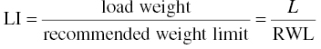
The revised NIOSH lifting equation yields a unique set of evaluation parameters that include (i) intermediate task-related multipliers that define the extent of physical stress associated with individual task factors; (ii) the NIOSH recommended weight limit (RWL), a task-specific value that defines the load weight that is considered safe for nearly all healthy workers; and (iii) the NIOSH lifting index (LI), which provides a relative estimate of the overall physical stress associated with a specific manual lifting task.
The RWL is defined by the following equation:
where the load constant (LC) is equal to 51 lb and the terms HM, VM, DM, AM, CM, and FM are task-specific multipliers within the equation that serve to reduce the recommended weight limit according to the specific task factor to which each multiplier applies. The magnitude of each multiplier will range in value between zero and one, depending on the value of the task factor to which the multiplier applies. The multipliers are defined as in Table 3.5.
TABLE 3.5 Formulas for individual multipliers.
| Component | Metric | US customary |
| HM = horizontal multiplier | (25/H) | (10/H) |
| VM = vertical multiplier | [1 − (0.003∣V − 75∣)] | [1 − (0.0075∣V − 30∣)] |
| DM = distance multiplier | [0.82 + (4.5/D)] | [0.82 + (1.8/D)] |
| AM = asymmetric multiplier | [1 − (0.0032A)] | [1 − (0.0032A)] |
| FM = frequency multiplier | (Determined from Table 3.6) | |
| CM = coupling multiplier | (Determined from Table 3.7) |
In order to use the revised NIOSH lifting equation, you need to make the following measurements:
- L = weight of the load being lifted (lb or kg).
- H = horizontal location of the hands from midpoint between the ankles. Measure at the origin and the destination of the lift (cm or in).
- V = vertical location of the hands above the floor. Measure at the origin and destination of the lift (cm or in).
- D = vertical travel distance between the origin and the destination of the lift (cm or in).
- A = angle of asymmetry, defined as the angular displacement of the load from the sagittal plane when lifts are made to the side of the body. Measure at the origin and destination of the lift (°).
- F = average frequency rate of lifting measured in lifts/minute. Duration is defined as <1, 1–2, or 2–8 hours. Specific recovery allowances are required for each duration category (see Tables 3.6 and 3.7).
TABLE 3.6 Frequency multiplier (FM).
Values of V are in cm; 75 cm = 30 in.
| Frequency lifts/minute | Work duration | |||||
| ≤1 hour | ≤2 hours | ≤8 hours | ||||
| V < 75 | V ≥ 75 | V < 75 | V ≥ 75 | V < 75 | V ≥ 75 | |
| 0.2 | 1.00 | 1.00 | 0.95 | 0.95 | 0.85 | 0.85 |
| 0.5 | 0.97 | 0.97 | 0.92 | 0.92 | 0.81 | 0.81 |
| 1 | 0.94 | 0.94 | 0.88 | 0.88 | 0.75 | 0.75 |
| 2 | 0.91 | 0.91 | 0.84 | 0.84 | 0.65 | 0.65 |
| 3 | 0.88 | 0.88 | 0.79 | 0.79 | 0.55 | 0.55 |
| 4 | 0.84 | 0.84 | 0.72 | 0.72 | 0.45 | 0.45 |
| 5 | 0.80 | 0.80 | 0.60 | 0.60 | 0.35 | 0.35 |
| 6 | 0.75 | 0.75 | 0.50 | 0.50 | 0.27 | 0.27 |
| 7 | 0.70 | 0.70 | 0.42 | 0.42 | 0.22 | 0.22 |
| 8 | 0.60 | 0.60 | 0.35 | 0.35 | 0.18 | 0.18 |
| 9 | 0.52 | 0.52 | 0.30 | 0.30 | 0.00 | 0.15 |
| 10 | 0.45 | 0.45 | 0.26 | 0.26 | 0.00 | 0.13 |
| 11 | 0.41 | 0.41 | 0.00 | 0.23 | 0.00 | 0.00 |
| 12 | 0.37 | 0.37 | 0.00 | 0.21 | 0.00 | 0.00 |
| 13 | 0.00 | 0.34 | 0.00 | 0.00 | 0.00 | 0.00 |
| 14 | 0.00 | 0.31 | 0.00 | 0.00 | 0.00 | 0.00 |
| 15 | 0.00 | 0.28 | 0.00 | 0.00 | 0.00 | 0.00 |
| >15 | 0.00 | 0.00 | 0.00 | 0.00 | 0.00 | 0.00 |
TABLE 3.7 Coupling multiplier (CM).
| Couplings | Coupling multipliers | |
| V < 75 cm (30 in.) | V ≥ 75 cm (30 in.) | |
| Good | 1.00 | 1.00 |
| Fair | 0.95 | 1.00 |
| Poor | 0.90 | 0.90 |
The LI provides a relative estimate of the level of physical stress associated with a particular manual lifting task. The estimate of the level of physical stress is defined by the relationship of the weight of the load lifted to the recommended weight limit. The LI is defined by the following equation:
A detailed explanation of the use of the revised NIOSH lifting equation, including definitions of terms and procedures, is available in an applications manual for the revised NIOSH lifting equation.64 The document can be downloaded from the NIOSH website (https://www.cdc.gov/niosh/docs/94-110/).
The developers of the revised NIOSH lifting equation indicated that studies were needed to determine the effectiveness of the NIOSH lifting equation in identifying jobs with increased risk of lifting-related LBP for workers. Researchers at NIOSH published the results of a cross-sectional epidemiologic study designed to evaluate the effectiveness of the equation in identifying jobs with elevated risk of causing LBP.65 In the NIOSH study, 50 jobs were evaluated using the revised NIOSH lifting equation. The LI values of the jobs were compared to information obtained about the LBP symptoms in people who worked in those jobs. Using logistic regression modeling, the odds ratio (OR) for LBP was determined for various categories of LI values compared to the unexposed control group. LBP was assessed with a symptom and occupational history questionnaire that was administered to 204 workers employed in lifting jobs and 80 workers employed in nonlifting jobs. Participation was 89–95% among the exposed workers and 82–100% among unexposed workers at four facilities. The authors found that as the LI increased from 1.0 to 3.0, the odds of LBP increased, with a peak and statistically significant OR occurring in the two to three LI category (unadjusted OR = 2.45; CI 1.29–4.85). For jobs with a LI higher than 3.0, however, the OR was lower (1.63; CI 0.66–3.95). The decrease in the OR for these highly exposed jobs is likely to result from a combination of worker selection and a survivor effect. This study also examined several confounding variables such as age, gender, body mass index, and psychosocial factors that were included in the multiple logistic models. The highest OR was in the two to three LI category (2.2; CI 1.01–4.96).
Additional studies conducted in the last decade have broadened the research base that has established the revised NIOSH lifting equation and confirmed its versatility as a valid methodology for estimating LBP risk. For jobs that require multiple MMH activities, a new index methodology has been developed. This method is based on the concept that a composite lifting index (CLI), which represents the collective demands of the job, is equal to the sum of the largest single task lifting index (STLI) and the incremental increases in the CLI as each subsequent task is added. The incremental increase in the CLI for a specific task is defined as the difference between the lifting index for that task at the cumulative frequency and the lifting index for that task at its actual frequency. Other studies have incorporated individual, psychosocial, and work organizational risk factors and their influence on self-reported back pain and the calculation of the lifting index. An excellent article which summarizes this recent research involving the revised NIOSH lifting equation is available from Lu et al.66
DIAGNOSIS AND TREATMENT
The primary objectives of the diagnosis and treatment of occupationally related musculoskeletal injuries are to (i) assist the recovery of workers and allow for a rapid return to work (ii) ensure that proper diagnostic tools are used so that an accurate assessment is made of the magnitude of the injury, and (iii) provide appropriate cost-effective treatments that avoid unnecessary surgery. The system should be based on an organized approach to evaluation, diagnosis, and treatment, which is essential for an early return to work and reduction in costs and lost work time. For example, a standardized diagnostic and treatment protocol has been shown to be effective in significantly and continuously reducing the number of incidents, days lost from work, low back surgery cases, and financial costs of LBP.67
Although a variety of MSDs result from MMH, the single most costly medical condition is work-related LBP, which affects millions of Americans. Experts have indicated that there is significant variation in assessment and treatment of LBP that results in inappropriate or at least less than optimal care for many patients with low back disorders. This issue was addressed in 1994 with the publication of the Clinical Practice Guideline for Acute Low Back Pain in Adults by the Agency for Health Care Policy and Research (AHCPR).68 Copies of the guidelines are available on the agency’s (now renamed the Agency for Healthcare Research and Quality) website at www.ahrq.gov. These guidelines, although somewhat outdated, focused on returning the patient to normal activity and were in widespread use. A more recent review, which is more inclusive and includes additional return to work (RTW) models, is provided by Schultz et al.69
Complicating the diagnosis and treatment of occupationally related LBP are the legal issues of disability and compensation and how they relate to pain and impairment. Pain and impairment, which are direct measures of the extent of injury, primarily depend on the severity of the injury. Disability and compensation, however, may depend more on the nature of the compensation system and laws than on the severity of the injury.70
Diagnosis
Acute low back pain is often nonspecific and it is often difficult to identify a specific cause. There are some estimates that more than 90% of all episodes of back pain are probably attributable to mechanical causes,71 which could certainly be the case with MMH activities. Thus, the early diagnostic evaluation of back pain is designed to rule out systemic disease; grossly identify neurologic or anatomic abnormalities that may eventually require surgery such as fracture, tumor, infection, or cauda equina syndrome; and identify characteristic indicators of injury that may influence the selection of treatment. Clinical guidelines for diagnosis and treatment of low back pain are available from Chou et al.72 and Casazza.73
The first step in diagnosing LBP, and perhaps the most important, is the medical history. The medical history should include an assessment of current symptoms, individual history of injury, functional status, and injury documentation and psychosocial risk factors which may be helpful in predicting risk for chronic back pain.72,74–77 The second step in the diagnostic process is a complete physical examination. At minimum, this examination would include a check of the deep tendon reflexes at the Achilles and quadriceps tendons, bilateral straight-leg raising, palpation of the paraspinal muscles for spasm, and a screening motor and sensory neurologic examination. The neurologic examination should give special attention to the L5 and S1 nerve roots, since the vast majority of disk herniations occur at either the L4–L5 or L5–S1 interspaces. It has been suggested that a system should be developed to ensure that all parts of the examination are completed.73,76
Chou et al. proposes that after the medical history and physical examination patients be categorized into three broad categories: (i) nonspecific low back pain, (ii) back pain potentially associated with radiculopathy or spinal stenosis, or (iii) back pain potentially associated with another specific spinal cause.72 Most back injuries from MMH activities would be categorized as nonspecific.
Imaging is not recommended for most patients without signs and symptoms of a serious condition.73 Magnetic resonance imaging (MRI) may be most appropriate if a serious condition is suspected. Computed tomography can be used if an MRI is unavailable. However, these sophisticated imaging tests should not be ordered too early in the course of back pain, especially when there is an absence of clinical findings that suggest a need for surgical intervention. In general, these tests should be limited to workers who have continued neurologic findings after 4–6 weeks of conservative therapy. It is also important to remember that an abnormal imaging study and back pain are not necessarily related. The clinical course of each worker must be considered before this causal link is made and surgery is considered.
Treatment
Epidemiologic data suggest that the vast majority of low back injuries are not serious and that most workers return to work after a short time with only conservative therapy. In general, only a minority of all affected persons have back pain >2 weeks in duration. By contrast, 90% of patients return to work within 6 weeks of onset of the back pain.76,78
Recent recommendations for treatment on nonspecific back pain have changed from previous recommendations as evaluations of effective treatments improved. Practitioners treating LBP should review the ACOEM Practice Guidelines for low back disorders.79
Casazza73 categorizes treatments into (i) recommended, (ii) acceptable, (iii) unsupported, and (iv) inadvisable, using an evidence-based approach. Recommended treatments are medications and patient education. Nonsteroidal anti-inflammatory drugs (NSAIDS) are effective for short-term relief as are benzodiazepine muscle relaxants. Opioids, while frequently prescribed, showed little evidence of benefit over NSAIDS in relief or return to work. Epidural steroid injections are frequently offered after 2–6 weeks of no relief from radicular pain with noninvasive treatment. However, the FDA has not approved corticosteroids for such use and has stated that the effectiveness and safety of the drugs for this use have not been established.80 Patient education involves advising patients to stay active as much as the pain allows, avoid twisting and bending, and return to normal activities as soon as possible. Assurance that acute back pain is mostly benign in nature is important. Acceptable treatments are physical therapy, especially the McKenzie method,81 and physical therapist-directed home exercise and application of ice or heat treatments in the first 5 days of pain occurrence. Unsupported treatments include oral steroids, acupuncture, exercise, lumbar support, massage, spinal manipulation, and traction. Many of the treatments in the unsupported list lack high-quality studies to evaluate intervention effectiveness but may be beneficial for some patients. An inadvisable treatment is bed rest, especially prolonged bed rest.
If conservative therapy fails and 4–6 weeks have passed, further diagnostic tests should be scheduled. These cases generally fall into one of the following four categories, by location of the patient’s predominant complaints: (i) LBP, which is typically diagnosed as back sprain; (ii) leg pain radiating below the knee, which is commonly referred to as sciatica; (iii) posterior thigh pain; and (iv) anterior thigh pain. Each of these conditions warrants a unique treatment regimen.67 It may also be appropriate to thoroughly investigate and address psychological and psychosocial issues that were not identified at the initial assessment. Failure to deal with a significant psychological or psychosocial issue will delay the patient’s return to work and may ultimately result in treatment failure and continuing disability.
A few back pain patients have symptoms suggesting sciatica, which is usually the first clue to a herniated disk. It is estimated that only 5–10% of patients with persistent sciatica require surgery. In general, only patients diagnosed with cauda equina compression (CEC) or a similar mechanoanatomic problem require immediate surgical intervention. Other indications for referral include severe or progressive neurologic deficits, or persistent neurologic finding after 4–6 weeks of conservative therapy. In the overwhelming number of cases of LBP, the appropriate treatment includes a course of conservative therapy.67,82
There is general agreement among clinicians that treatment for LBP must be given according to a strict timetable so that patients do not develop a dependence that would prolong symptoms and functional limitations. Also, patient reassurance and education is an important aspect of therapy. The clinician must avoid labeling the patient. The patient should be reassured that the natural history of their condition is favorable and that he or she should be able to return to work in a short time.82
MEDICAL SURVEILLANCE
Active and passive surveillance of the workplace are necessary to establish a database of potential exposures in each workplace. Medical management of injuries and early reporting should be incorporated within the general medical treatment program at each work site. Since there is usually a short latency period between exposure and the onset of symptoms, medical surveillance per se is not helpful in eliminating MMH injuries. It may be useful to screen employees in order to match their physical abilities to the job. However, this is more of an exercise in prevention of injuries and is thus covered in the next section on “Prevention.”
PREVENTION
Several strategies have been developed to try and prevent MSD injuries from MMH activities. These strategies can be categorized into three general types: (i) workplace directed, (ii) worker directed, and, (iii) employee screening.
Workplace-directed approaches
AUTOMATION
Workplace automation should be a top priority when the job has high physical demands, is highly repetitive, or is performed in a hazardous environment. Automation may consist of one or more machines or machine systems such as conveyors, automated handling lines, automated storage and retrieval systems, or robots. This approach is best suited for the design of new work processes or activities or for the redesign of highly stressful tasks. Because automation often requires large capital expenditures, this approach may be prohibitive for small companies with only a few workers.
MECHANICAL AIDS
In cases where the physical demands are high and automation is not practical, mechanical aids can be used to ameliorate the extent of those demands. Mechanical handling aids include machines or simple devices that provide a mechanical advantage during the MMH task, such as hand trucks, cranes, hoists, lift tables, powered mobile equipment and lift trucks, overhead handling and lifting equipment, and vacuum lift devices. As mentioned earlier, the publication Ergonomic Guidelines for Manual Material Handling illustrates a number of mechanical aids that would be useful in reducing exposure to MMH.2 Additionally, a recent NIOSH publication Ergonomic Solutions for Retailers is specifically oriented for prevention of MMH injuries in the grocery sector and illustrates mechanical aids that are useful in the grocery sector.83
ERGONOMIC DESIGN CHANGES
For MMH jobs, ergonomic design or redesign may be accomplished by modifying the job layout, engineering redesign, or incorporating procedures to reduce bending, twisting, horizontal extensions, heavy lifting, forceful exertions, and repetitive motions
. The ergonomic approach is largely based on the assumption that work activities involving less weight, repetition, awkward postures, and applied force are less likely to cause injuries and disorders. The ergonomic approach is desirable because it seeks to eliminate potential sources of problems. Ergonomics also seeks to make safe work practices a natural result of the tool and work site design.
There are at least four advantages to adopting an ergonomic design/redesign strategy. First, an ergonomic approach does not depend on specific worker capabilities or learned behaviors, such as training. Second, human biological factors and their variations are accounted for in ergonomic approaches using design data that accommodate large segments of the populations. Third, ergonomic intervention is relatively permanent, since the workplace hazard is eliminated. Fourth, to the extent that sources of biomechanical stress at the work site are eliminated or significantly reduced, the difficult issues involving potential worker discrimination, lifestyle modifications, or attempts at changing behavioral patterns of workers will be of lesser practical significance. Many MMH jobs entail a variety of specialized tasks with overlapping sources of physical stress. Each MMH task may contribute in some unknown manner to the onset of an overexertion injury. Often, no single job modification is feasible or sufficient, and numerous adjustments may be required for each activity to minimize the hazard of overexertion injury. It is always important to develop an evaluation process to determine if the job modification is effective.
Worker-directed approaches
TRAINING AND EDUCATION
According to OSHA,84 the purpose of training and education is to “ensure that employees are sufficiently informed about the ergonomic hazards to which they may be exposed and thus are able to participate actively in their own protection.” Training may include general instruction on the types of injuries that may occur, what risk factors may contribute to these injuries, how to recognize and report symptoms, and how to prevent these injuries. Training should also include job-specific instruction on the proper use of essential MMH equipment and techniques for the specific task or job. The training program should include the following individuals: (i) all exposed workers, (ii) supervisors, (iii) managers, (iv) engineers and maintenance personnel, and (v) healthcare providers.
The term training has been used to describe two distinctly different approaches to injury prevention and control—instructional training in safe materials handling and fitness training (e.g., conditioning, strengthening, or work hardening). The basic premise of instructional training in MMH is that people can more safely handle greater loads when they perform the task correctly than if they perform the task incorrectly. Fitness training is based on the premise that people can safely handle greater loads when their strength or aerobic capacity is increased.
INSTRUCTIONAL TRAINING
Instructional training is widely used and fundamentally sound, but there some potential problems when not appropriately used. First, care must be taken to ensure that appropriate, scientifically sound work practices are being taught. Today, with the advent of the Internet and the mass media exposure, work practices that have not been properly evaluated can be promoted that may be entirely unsound for MMH activities. For example, back belts were introduced as a device for preventing back injuries and were implemented in many workplaces with instructional techniques before scientific evaluation showed limited usefulness. NIOSH concluded, in 1994, that there was insufficient evidence to recommend the use of back belts as a back injury prevention measure.85,86 Since then, NIOSH conducted a large epidemiologic study and two laboratory evaluations to determine more conclusively the effects of back belt use. The further research did not provide evidence to change NIOSH’s earlier conclusions.87–89
Additionally, in some training courses, workers are taught to lift with a straight back and bent legs (i.e., squat posture) rather than bending over to pick up the load (i.e., stooped posture). This either/or approach does not always provide the “safest” lifting style.62 Recent research has shown that the horizontal distance of the load from the worker may be more important in determining the spinal loading forces that the amount of forward bending. For this reason, NIOSH does not recommend a single, correct lifting style for all manual lifting tasks, but suggests that a free style lift, in which workers choose whatever style they prefer may be the most appropriate keeping in mind the avoidance of excessive bending and reach.
Secondly, the instructional approach relies on the worker’s ability to comply with a set of recommended practices that may be forgotten or changed from time to time. To some extent, worker’s ability to comply with safe practices can be improved with training materials that are more specifically directed at the MMH activities they are involved with. Two recent NIOSH documents such as Simple Solutions for Home Building Workers90 and the aforementioned Ergonomic Solutions for Retailers are examples.83
Regardless of the potential problems associated with MMH training, all workers who perform MMH activities should receive basic instructional training in the recognition of hazardous tasks and should have a thorough knowledge of what to do when a hazardous task is identified. Furthermore, the instructional training should provide information to workers on how they can become involved in the process of preventing and controlling injuries on the job. An excellent review of the effectiveness of training and education programs for prevention of workplace injuries has recently been published.91
FITNESS TRAINING
The basic premise of fitness training is that a worker’s risk of injury would decrease if his or her strength or fitness increased. Although this seems intuitive, it is not clear whether an individual’s strength and risk of injury performing MMH activities are related. Certainly, a worker’s capacity to perform heavy work might be increased, but there is some controversy about the relationship between worker strength and risk of injury.92 Moreover, it is not known how the soft tissues of the body respond to increased loads associated with stronger muscles, especially if the worker performs tasks requiring greater strength demands. Regardless of strength, an improper lifting technique could lead to an injury. Strength and fitness exercises are available in several documents and in preparation for MMH activities.90
Although both types of training programs have been used to prevent MMH injuries, the effectiveness of training in preventing or controlling injuries is unclear at the present time. Therefore, training programs should be used as a supplement to workplace-directed approaches.
Employee screening
Screening methodologies, which rely on the assessment of one or more physical characteristics of the worker to select specific workers for certain MMH jobs, have been advocated by some ergonomic experts. These methodologies are designed to identify workers (i) with a high risk of overexertion injury, such a history of previous physical injury that may limit MMH activities, and/or (ii) screen workers according to some preselected set of strength or endurance criteria in an attempt to match the capacity of the worker to the demands of the job.
RISK ASSESSMENT SCREENING
Attempts to identify workers with a high risk of overexertion injury have included such activities as spinal radiographs, psychological testing, and medical examinations, which are designed to provide an objective basis for excluding certain individuals from stressful MMH jobs. Few of these methodologies, however, have been shown to be reliable in predicting an individual’s risk of overexertion injury. Although no psychological tests have been found to quantify a worker’s risk of overexertion injury, psychological testing can provide an indication of how a worker might respond to a severe injury. Shaw et al. evaluated patients with recent onset of occupational low back pain and using a survey of potential disability risks and a questionnaire reporting symptoms of pain.93 Return to work was more strongly related to employer factors (e.g., job tenure, physical work demands, and modified duty availability) and self-ratings of pain and mood than by health history or physical examination.
PHYSICAL CAPACITY SCREENING
Another type of screening approach that has been used to select workers for MMH tasks includes individual testing of physical characteristics, such as strength, aerobic capacity, or functional capability. The underlying basis for using tests such as these to screen workers for MMH jobs is the belief that the risk of injury is dependent on the relationship between the capacity of the worker and the demands of the job. When the physical demands of the job exceed the capacity of the worker, the worker is at risk for developing an MSD. Thus, the idea of this approach is that workers should be matched to jobs according to the demands of the work.
A number of studies have been conducted to develop databases of maximum strength capacities (i.e., population averages) that could be used to design MMH tasks and workstations. These studies, however, disagree as to which of the three principal testing methods—isometric, isokinetic, or isoinertial—is most useful for determining strength capacity guidelines. Some researchers argue that traditional isometric lifting strength measurements, by which thousands of workers have been tested, are limited in assessing what workers can do under dynamic task conditions. These researchers suggest that dynamic strength testing is more appropriate than static strength testing for determining strength capacity.94 This assertion is based on how well the test replicates the job requirements. Other researchers, however, claim that isokinetic lifting strength measurements probably have no greater inferential power to predict risk of injury or job performance than any other form of testing.95 Kroemer claims that isoinertial strength testing is the most appropriate lifting strength testing method because it matches actual lifting conditions.96 Isoinertial methods, however, have not been generally validated in terms of their ability to predict risk of injury.97
Maximum isometric lifting strength (MILS) has been studied and reported extensively. It has a well-established testing procedure98 and has been reported in field tests to predict risk of injury.22,99 Extensive measures of MILS have been made for various work postures and activities. One study, for example, measured the isometric strength of 1239 workers in rubber, aluminum, and electronic component industries.100 In another study that measured the standardized isometric strengths (i.e., arm lift, torso lift, and leg lift) of 2178 aircraft manufacturing workers,90 the employees were followed for >4 years to document back pain complaints. The investigators found that worker height, weight, age, and gender are poor predictors of standardized isometric strength, a finding that agrees with other studies.100 The investigators also found, however, that standardized isometric strength is a poor predictor of reported back pain, a finding that conflicts with the results of some other studies.101
Marras et al. published an ergonomic guide for assessing dynamic measures of low back performance.102 This guide provides information on elements of dynamic performance, techniques to assess dynamic performance, and relationships between testing techniques and internal forces. Bos et al. reported the results of a systematic literature search to identify specific lifting, pushing, and pulling demands and evaluate reliable and valid tests for the assessment of acceptable load on an individual level.103 Results showed that there was no universal strategy for the definition of occupational demand and none of the tests met the criteria of reliability and prognostic value for musculoskeletal complaints completely on an individual level. There was some epidemiological evidence that for the development of musculoskeletal complaints, there was prognostic value with strength capacity tests, but not for a maximum acceptable load at an individual level.
SUMMARY
In summary, MMH activities, such as excessive lifting, pushing, pulling, and carrying, represent a serious hazard for MSD for many workers. There are analytic tools to identify ergonomic hazards that may result in overexertion injury and prevention tools that are effective in reducing the potential for risk of work-related overexertion injury. Successful ergonomic programs require the full cooperation of management, labor organizations, government, and the workers themselves. The solution requires a team effort with a commitment to identify and eliminate hazardous material handling tasks from the workplace.
Current status and future concerns
There is clear evidence that many MMH activities that involve lifting, pushing, pulling, or carrying present risks to workers for developing MSDs. In the past MSDs were frequently just considered as a cost of doing business, but as health problems mounted coupled with the increased costs to employers and taxpayers to deal with these increases, attitudes have changed. Prevention of MSDs from MMH activities has come to the forefront.
In this chapter MMH activities and the potential MSDs that can result have been described, and strategies for preventing and identifying MSDs have been presented. Analytical tools have been presented to identify the physical hazards that lead to injuries and are effective in reducing the risks for developing work-related MSDs.
Evident in this chapter is that much knowledge has been gained in the assessment and treatment of MSDs related to MMH activities; there are also significant research gaps that also exist. Several of these gaps have been identified by national expert reviews.11,12,104 A list of some identified research gaps is presented below.
- How accurate are methods for measuring a worker’s maximum safe capacity? Worker selection approaches assume that it is possible to accurately determine a worker’s safe capacity. In addition, does the safe capacity change over time (e.g., hourly, daily, monthly, yearly)?
- Ergonomic approaches, which are considered to be more effective in reducing MSDs, can also be expensive to implement. Do such approaches have reasonable cost/benefit advantages?
- How accurate are the existing assessment tools (e.g., biomechanical, psychophysical, physiological, integrated) that are used to determine risks of injury from MMH tasks. Do these tools have limitations in predicting long-term risks?
- Do performance standards or incentive programs increase a workers’ risk of injury from MMH activities? If performance standards are used, are there criteria available to ensure safety for the worker?
- What are the effects psychosocial risk factors (job stress, organizational, job safety, work time, work freedom, work schedule) on MMH activities that can influence the risk of MSDs?
- The workforce is changing, with older workers and more women involved in MMH activities that require frequent lifting, pushing, and pulling. Are these changes being incorporated into job designs that can reduce the physical demands of many MMH activities?
These questions, although not exhaustive, identify the many issues that safety and health experts and industry leaders must address to reduce work-related injuries. Efforts to limit workplace hazards will only be successful with the full cooperation of management, government, and labor and, of course, the workers themselves. Solutions will require collaborative efforts of all involved.
References
- 1. International Organization for Standardization. ISO 11228-1:2003. Ergonomics: Manual Handling. Part 1: Lifting and Carrying. Geneva: ISO, 2003.
- 2. Cal/OSHA Consultation Service and the National Institute for Occupational Safety and Health. Ergonomic Guidelines for Manual Material Handling. DHHS (NIOSH) Publication No. 2007-131. Sacramento/Cincinnati, OH: Department of Industrial Relations/Department of Health and Human Services, Centers for Disease Control and Prevention, National Institute for Occupational Safety and Health, 2007.
- 3. Liberty Mutual Research Institute for Safety. 2014 Liberty Mutual Workplace Safety Index. Hopkinton, MA: Liberty Mutual Research Institute for Safety, 2014.
- 4. Centers for Disease Control and Prevention. Nonfatal Work-Related Injuries and Illnesses – United States, 2010. CDC Health Disparities and Inequalities Report—United States, 2013. MMWR 2013; 62 (Suppl 3):35–40.
- 5. Bureau of Labor Statistics. Nonfatal Occupational Injuries and Illnesses Requiring Days Away From Work, 2013. U.S. Department of Labor, USDL-14-2246, December 16, 2014.
- 6. National Safety Council. Injury Facts®. Itasca, IL: National Safety Council, 2015.
- 7. Waters TR, Dick RB, Davis-Barkley J, et al. A cross-sectional study of risk factors for musculoskeletal symptoms in the workplace using data from the general social survey (GSS). J Occup Environ Med 2007; 49(2):172–84.
- 8. Waters TR, Dick RB, Krieg EF. Trends in work-related musculoskeletal disorders: a comparison of risk factors for symptoms using quality of work life data from the 2002 and 2006 general social survey. J Occup Environ Med 2011; 53(9):1013–24.
- 9. Dick RD, Lowe BD, Lu M-L, et al. Further trends on work-related musculoskeletal disorders: a comparison of risk factors for symptoms using quality of work life data from the 2002, 2006 and 2010 general social survey. J Occup Environ Med 2015; 57(8):910–28.
- 10. Bogduk N. Clinical Anatomy of the Lumbar Spine and Sacrum, 5th ed. New York: Elsevier/Churchill Livingstone, 2012.
- 11. National Institute for Occupational Safety and Health. Musculoskeletal Disorders and Workplace Factors: A Critical Review of Epidemiological Evidence for Work-Related Musculoskeletal Disorders of the Neck, Upper Extremity, and Low Back. DHHS (NIOSH) Publication No. 97-141. Washington, DC: US Department of Health and Human Services, 1997.
- 12. National Research Council, Institute of Medicine—Panel on Musculoskeletal Disorders and the Workplace, Commission on Behavioral and Social Sciences and Education. Musculoskeletal Disorders and the Workplace: Low Back and Upper Extremities, Washington, DC: National Academy Press, 2001.
- 13. da Costa BR, Vieira RE. Risk factors for work-related musculoskeletal disorders: a systematic review of recent longitudinal studies. Am J Ind Med 2010; 53:285–323.
- 14. Pope MH, Frymoyer JW, Andersson G. Occupational Low Back Pain. New York: Praeger, 1984.
- 15. Ferguson SA, Marras WS. A literature review of low back disorder surveillance measures and risk factors. Clin Biomech 1997; 12(4):211–26.
- 16. Mayer J, Kraus T, Ochsmann E. Longitudinal evidence for the association between work-related physical exposures and neck and/or shoulder complaints: a systematic review. Int Arch Occup Environ Health 2012; 85:587–603.
- 17. Gallagher S, Heberger JR. Examining the interaction of force and repetition on musculoskeletal disorder risk: a systematic literature review. Hum Factors 2013; 55(1):108–24.
- 18. U.S. Department of Labor. Safety and Health Management Systems eTool website. Occupational Safety and Health Administration, http://www.osha.gov/SLTC/etools/safetyhealth/comp2.html accessed August 7, 2015.
- 19. David GC. Ergonomic methods for assessing exposure to risk factors for work-related musculoskeletal disorders. Occup Med 2005; 55:190–9. doi:1093/occmed/kqi082
- 20. van der Beek A, Frings-Dresen MHW. Assessment of mechanical exposure in ergonomic epidemiology. Occup Environ Med 1998; 55:190–5.
- 21. Dempsey PG, McGorry RW, Maynard WS. A survey of tools and methods used by certified professional ergonomists. Appl Ergon; 2005, 36:489–503.
- 22. Chaffin DB, Park KS. A longitudinal study of low-back pain as associated with occupational weight lifting factors. Am Ind Hyg Assoc J 1973; 34:513–25.
- 23. Frymoyer JW, Pope MH, Costanza MC, et al. Epidemiologic studies of low back pain. Spine 1980; 5:419–23.
- 24. Snook SH. The design of manual-handling tasks. Ergonomics 1978; 21:963–85.
- 25. Waters TR, Putz-Anderson V, Garg A, et al. Revised NIOSH equation for the design and evaluation of manual lifting tasks. Ergonomics 1993; 36(7):749–76.
- 26. The Eastman Kodak Company. Kodak’s Ergonomic Design for People at Work, 2nd ed. Hoboken, NJ: John Wiley & Sons, Inc., 2004.
- 27. Ayoub MM, Mital A, Asfour SS, et al. Review, evaluation, and comparison of models for predicting lifting capacity. Hum Factors 1980; 22(3):257–69.
- 28. Chaffin DB, Andersson GBJ, Martin BJ. Occupa-tional Biomechanics, 4th ed. New York: Wiley-Interscience, 2006.
- 29. Roderick D, Karwowski W. Manual materials handling. In: Salvendy G, ed. Handbook of Human Factors and Ergonomics, 3rd ed. Hoboken, NJ: John Wiley & Sons, Inc., 2006, pp. 818–54.
- 30. Chaffin DB, Baker WH. Biomechanical model for analysis of symmetric sagittal plane lifting. AIIE Trans 1970; 2(1):16–27.
- 31. Schultz AB, Andersson GBJ. Analysis of loads on the lumbar spine. Spine 1981; 6(1):76–82.
- 32. Freivalds A, Chaffin DB, Garg A, et al. A dynamic biomechanical evaluation of lifting maximum acceptable loads. J Biomech, 1984; 17(4):251–62.
- 33. McGill SM, Norman RW. Dynamically and statically determined low back moments during lifting, J Biomech 1985; 18(12):877–85.
- 34. Marras WS Sommerich CM. A three-dimensional motion model of loads on the lumbar spine: I. Model structure. Hum Factors 1991; 33(2):123–37.
- 35. Marras WS, Sommerich CM. A three-dimensional motion model of Loads on the lumbar spine: II. Model validation. Hum Factors 1991; 33(2):139–49.
- 36. Marras WS, Allread WG, Burr DL, et al. Prospective validation of a low-back disorder risk model and assessment of ergonomic interventions associated with manual materials handling tasks. Ergonomics 2000; 43(11):1866–86.
- 37. Kerr MS, Frank JW, Shannon HS, et al. Biomechanical and psychosocial risk factors for low back pain at work. Am J Pub Health 2001; 91(7):1069–75.
- 38. Skotte JH, Essendrop M, Hansen AF, et al. A dynamic 3D biomechnical evaluation on the load on the low back during different patient-handling tasks. J Biomech 2002; 35:1357–66.
- 39. Brinckmann P, Johannleweling N, Hilweg D, et al. Fatigue fracture of human lumbar vertebrae: brief report. Clin Biomech 1987; 2(2):94–6.
- 40. Stevens SS. Mathematics, measurement, and psychophysics. In: Stevens SS, ed. Handbook of Experimental Psychology. New York: John Wiley & Sons, Inc., 1951.
- 41. Ayoub MM. Psychophysical Basis for Manual Lifting Guidelines. Purchase order number 88-79313, 1988, Preliminary report received 12/16/88. Final report received 2/1/89. Cincinnati, OH: National Institute for Occupational Safety and Health, 1988.
- 42. Snook SH, Ciriello VM. The design of manual-handling tasks: revised tables of maximum acceptable weights and forces. Ergonomics 1991; 34:1197–213.
- 43. Ayoub MM, Mital A. Manual Materials Handling. London: Taylor & Francis, 1989.
- 44. Borg G. Psychophysical scaling with applications in physical work and the perception of exertion. Scand J Work Environ Health 1990; 16(Suppl 1):55–8.
- 45. Corlett EN, Bishop RP. A technique for assessing postural discomfort. Ergonomics, 1976; 19:175–82.
- 46. Rodgers SH, Yates JW, Garg A. The Physiological Basis for Manual Lifting Guidelines. Report No. 91-227-330. Springfield, VA: National Technical Information Service, 1991.
- 47. Garg A. A Metabolic Rate Prediction Model for Manual Materials Handling Jobs. Ann Arbor, MI: University of Michigan, 1976.
- 48. Astrand PO, Rodahl K. Textbook of Work Physiology, 3rd ed., New York: McGraw-Hill, 1986.
- 49. Ayoub MM. Determination and Modeling of Lifting Capacity. Final report. Grant no. 5-ROI-OH-00545002. Cincinnati, OH: Department of Health and Human Services, National Institute for Occupational Safety and Health, 1978.
- 50. Takala EP, Pehkonen I, Forsman M, et al. Systematic evaluation of observational methods assessing biomechanical exposures at work. Scand J Work Environ Health 2010; 36(1):3–24.
- 51. University of Wisconsin-Madison, Multimedia Video Task Analysis™ (MVTA™). Madison, WI: Ergonomics Analysis and Design Consortium. http://mvta.engr.wisc.edu accessed April 27, 2016.
- 52. University of Michigan, 3D Static Strength Prediction Program™ (3DSSPP™). Ann Arbor, MI: Center for Ergonomics. http://c4e.engin.umich.edu/tools-services/3dsspp-software/ accessed April 27, 2016.
- 53. Liu Y, Zhang Z, Chaffin D. Perception and visualization of human information for computer-aided ergonomic analysis. Ergonomics 1997; 40:818–33.
- 54. Lu M, Waters T, Werren D. Development of human posture simulation method for assessing posture angles and spinal loads. Hum Factors Ergon Manuf Serv Ind 2015; 25(1):123–36.
- 55. Lowe B. Accuracy of validity of observational estimated of wrist and forearm posture. Ergonomics 2004; 47(5):527–54.
- 56. Bao S, Howard N, Spielholz P, et al. Interrater reliability of posture observations. Hum Factors 2009; 51(3):292–309.
- 57. Marras WS, Fattalah F. Accuracy of three dimensional lumbar motion monitor for recording dynamic trunk motion characteristics. Int J Ind Ergon 1992; 9:75–87.
- 58. Marras WS, Schoenmarklin RW. Wrist motions in industry. Ergonomics 1993; 36:341–51.
- 59. Grant KA, Galinsky, TL, Johnson PW. Use of the actigraph for objective quantification of hand/wrist activity in repetitive work. In: Proceedings of the Human Factors and Ergonomics Society 37th Annual Meeting, Sheraton Seattle Hotel & Towers/Washington State Convention Center, October 11–15, 1993, Seattle WA. Santa Monica, CA: Human Factors and Ergonomics Society, pp. 720–4.
- 60. Roetenberg D, Slycke P. Xsens MVN: Full 6 DOF Human Motion Tracking using Miniature Inertial Sensors. Enschede: Xsens Technologies, 2013.
- 61. Fabers G, Kingma I, Bruijin S, et al. Optimal inertial sensor location for ambulatory measurement of trunk inclination. J Biomech 2009; 42:2406–9.
- 62. Garg A. What basis exists for training workers in correct lifting technique? In: Marras WS, Karwowski W, Smith JC, et al., eds. The Ergonomics of Manual Work. London: Taylor & Francis, 1993.
- 63. National Institute for Occupational Safety and Health. Work Practices Guide for Manual Lifting. NIOSH Technical Report No. 81-122. Cincinnati, OH: Department of Health and Human Services, National Institute for Occupational Safety and Health, 1981.
- 64. Waters TR, Putz-Anderson V, Garg A. Applications Manual for the Revised NIOSH Lifting Equation. DHHS (NIOSH) Publication No. 94-110. Cincinnati, OH: National Institute for Occupational Safety and Health, 1994.
- 65. Waters TR, Baron SL, Piacitelli LA, et al. Evaluation of the revised NIOSH lifting equation: a cross-sectional epidemiological study. Spine 1999; 24(4):386–95.
- 66. Lu M-L, Putz-Anderson V, Garg A, et al. Evaluation of the impact of the revised National Institute for Occupational Safety and Health Lifting Equation. Hum Factors 2016; 58(5):667–82.
- 67. Boden SD, Wiesel SW. Standardized approaches to the diagnosis and treatment of low back pain and multiply operated low back patients. In: Wiesel SW, Wiesel SW, eds. Industrial Low Back Pain: A Comprehensive Approach, 2nd ed. Charlottesville, VA: The Michie Company, 1989.
- 68. Bigos SJ, Bowyer OR, Braen RG, et al.. Acute Low Back Problems in Adults. Clinical Practice Guideline No. 14. AHCPR Publication No. 95-0642. Rockville, MD: Agency for Health Care Policy and Research, Public Health Service, U.S. Department of Health and Human Services, 1994.
- 69. Schultz IZ, Stowell AW, Feuerstein M, et al. Models of return to work for musculoskeletal disorders. J Occup Rehabil 2007; 17:327–52.
- 70. Andersson GBJ, Pope MH, Frymoyer JW, et al.. Epidemiology and cost. In: Pope MH, Andersson GBJ, Frymoyer JW, et al., eds. Occupational Low Back Pain: Assessment, Treatment, and Prevention. St. Louis, MO: Mosby-Year Book, 1991, pp. 95–113.
- 71. White AA, Gordon SL. Synopsis: workshop on idiopathic low back pain. Spine 1982; 7:141–9.
- 72. Chou R, Qaseem A, Snow V, et al. Diagnosis and treatment of low back pain: a joint clinical practice guideline from the American College of Physicians and the American Pain Society. Ann Intern Med 2007; 147(7):478–91.
- 73. Casazza BA. Diagnosis and treatment of acute low back pain. Am Fam Physician 2012; 85(4):343–50.
- 74. Silverstein BA, Fine U. Evaluation of Upper Extremity and Low Back Cumulative Trauma Disorders: A Screening Manual. Ann Arbor, MI: University of Michigan, School of Public Health, Occupational Health Program, 1984.
- 75. Frymoyer JW, Haldermans S. Evaluation of the worker with low back pain. In: Pope MH, Andersson GBJ, Frymoyer JW, et al., eds. Occupational Low Back Pain: Assessment, Treatment, and Prevention. St. Louis, MO: Mosby-Year Book, 1991, pp. 151–82.
- 76. Andersson GBJ, Frymoyer JW. Treatment of the acutely injured worker. In: Pope MH, Andersson GBJ, Frymoyer JW, et al., eds. Occupational Low Back Pain: Assessment, Treatment, and Prevention. St Louis, MO: Mosby-Year Book, 1991, pp. 183–94.
- 77. Putz-Anderson V. Cumulative Trauma Disorders: A Manual for Musculoskeletal Disorders of the Upper Limbs. London: Taylor & Francis, 1988.
- 78. Deyo RA. Non-operative treatment of low back disorders. In: Frymoyer JW, ed. The Adult Spine: Principles and Practice. New York: Raven, 1991.
- 79. Low back disorders. In: Hegmann KT, eds. Occupational Medicine Practice Guidelines. Evaluation and Management of Common Health Problems and Functional Recovery in Workers, 3rd ed. Elk Grove Village, IL: American College of Occupational and Environmental Medicine (ACOEM), 2011, pp. 333–796. http://www.guideline.gov/content.aspx?id=38438 accessed April 27, 2016.
- 80. Food and Drug Administration. FDA Drug Safety Communication: FDA Requires Label Changes to Warn of Rare but Serious Neurologic Problems after Epidural Corticosteroid Injections for Pain April 23, 2013. http://www.fda.gov/Drugs/DrugSafety/ucm394280.htm accessed April 27, 2016.
- 81. Machado L, de Souza M, Ferreira P, et al. The McKenzie method for low back pain. Spine 2006, 31(9):254–62.
- 82. Deyo RA, Loeser JD, Bigos SJ. Herniated lumbar intervertebral disk. Ann Intern Med 1990; 112:598–603.
- 83. Putz-Anderson V, NIOSH. Ergonomic Solutions for Retailers: Prevention of Material Handling Injuries in the Grocery Sector. DHHS (NIOSH) Publication No. 2015-100. Cincinnati, OH: U.S. Department of Health and Human Services, Centers for Disease Control and Prevention, National Institute for Occupational Safety and Health, 2014.
- 84. Occupational Safety and Health Administration. Ergonomics Program Management Guidelines for Meatpacking Plants. OSHA Document No. 3123. Washington, DC: Department of Labor, Occupational Safety and Health Administration, 1990.
- 85. NIOSH. Workplace Use of Back Belts. DHHS (NIOSH) Publication No. 94-122. Cincinnati, OH: U.S. Department of Health and Human Services, Centers for Disease Control and Prevention, National Institute for Occupational Safety and Health, 1994.
- 86. NIOSH. Back Belts: Do They Prevent Injury? DHHS (NIOSH) Publication No. 94-127, Cincinnati, OH: U.S. Department of Health and Human Services, Centers for Disease Control and Prevention, National Institute for Occupational Safety and Health, 1994.
- 87. Wassell JT, Gardner LI, Landsittel DP, et al. A prospective study of back belts for prevention of back pain and injury. JAMA 2000; 284(21):2727–32.
- 88. Bobick TG, Belard J-L, Hsaio H, et al. Physiological effects of back belt wearing during asymmetric lifting. Appl Ergon 2001; 32:541–7.
- 89. Giocelli RJ, Hughes RE, Wassell JT, et al. The effect of wearing a back belt on spine kinematics during asymmetric lifting of large and small boxes. Spine 2001; 26(16):1794–8.
- 90. NIOSH. Simple Solutions for Home Building Workers: A Basic Guide for Preventing Manual Material Handling Injuries. DHHS (NIOSH) Publication No. 2013-111. Cincinnati, OH: U.S. Department of Health and Human Services, Centers for Disease Control and Prevention, National Institute for Occupational Safety and Health, 2013.
- 91. Robson L, Stephenson C, Schulte P, et al. A Systematic Review of the Effectiveness of Training and Education for the Protection of Workers. DHHS (NIOSH) Publication No. 2010-127. Toronto/Cincinnati, OH: Institute for Work & Health/National Institute for Occupational Safety and Health, 2010.
- 92. Battíe MC, Bigos SJ, Fisher LD, et al. Isometric lifting strength as a predictor of industrial back pain reports. Spine 1989; 14(8):851–6.
- 93. Shaw WS, Pransky G, Patterson W, et al. Early disability factors for low back pain assessed at outpatient health clinics. Spine 2005; 30(5):572–80.
- 94. Kroemer KHE. Testing individual capability to lift material: repeatability of a dynamic test compared with static testing. J Safety Res 1985; 3:4–7.
- 95. Rothstein JM, Lamb RL, Mayhew TP. Clinical uses of isokinetic measurements. Phys Ther 1987; 67:1840–4.
- 96. Kroemer KHE. An isoinertial technique to assess individual lifting capacity. Hum Factors 1983; 25:493–506.
- 97. Kroemer KHE. Matching individuals to the job can reduce manual labor injuries. Occup Safety Health News Dig 1987; 3:4–7.
- 98. Chaffin DB. Ergonomics guide for the assessment of human static strength. Am Ind Hyg Assoc J 1975; 36:505–11.
- 99. Herrin GD, Jaraiedi M, Anderson CK. Prediction of overexertion injuries using biomechanical and psychophysical modes. AIHA J 1986; 47:322–30.
- 100. Herrin GD, Keyserling WM, Chaffin DB, et al. A Comparison of selected work muscle strength. In: Proceeding of the 22nd Annual Meeting of the Human Factors Society, October 1978, Detroit, MI. Santa Monica, CA: Human Factors and Ergonomics Society.
- 101. Keyserling WM, Herrin GD, Chaffin DB. Isometric strength testing as a means of controlling medical incidents on strenuous jobs. J Occup Med 1980; 22:332–6.
- 102. Marras WS, McGlothlin JD, McIntyre DR, et al. Dynamic Measures of Low Back Performance: An Ergonomics Guide. Fairfax, VA: American Industrial Hygiene Association, 1993.
- 103. Bos J, Kuijer PPFM, Frings-Dresen MHW. Definition and assessment of specific occupational demands concerning lifting, pushing, pulling based on systematic literature search. Occup Environ Med 2002; 59:800–6.
- 104. Marras WS, Cutlip RG, Burt SE, et al. National occupational research agenda (NORA) future direction in occupational musculoskeletal disorder health research, Appl Ergon 2009; 40:15–22.