Introduction
From Conventional Single-phase Microfluidics to Droplets and Digital Microfluidics
Starting in the year 1980, microfluidics was at first a mere downscaling of macrofluidics. Its development was triggered by the emergence of biotechnology and materials science, imagined by visionary pioneers like Feynman [1], deGennes [2], Whitesides [3] and others. In particular, biotechnology was as a new science at the boundary of physics and biology. The goal was to give biological, medical and pharmaceutical research new automation tools to boost the development of new drugs, fabricate new body implants and increase the potentialities of fundamental research. In reality, this plan imagined by these first researchers has been extremely effective and produced even more discoveries than what was first expected. In a way, biotechnology developments bloomed according to Feynman’s words: “The best way to predict the future is to invent it.” The foreseen goals have required the downscaling of fluidic systems to the “convenient” size to work at the proper scale characteristic of a population of biologic targets. At the same time, it was found that the downscaling brought economy in costly materials, fluids, and devices; that sensitivity was increased and operating times were greatly reduced by the integration of many functions on the same microchip. Gradually, as microsystems based on microflows become conventionally used, new approaches were investigated that required even less volume of sample fluids. This trend to downscaling has promoted the development of new microfluidic approaches such as droplet and digital microfluidics. Reduction of the liquid vessel containing the biological targets was found to be possible by the use of microdroplets. New systems based on the confinement of biologic targets in extremely small vessels like microdrops are emerging. In such approaches the liquid volumes are reduced to a few picoliters.
Domains of Application
Historically, genomics and proteomics were the first beneficiaries of the development of biotechnology, and now it is the turn of cellomics. Also, these developments have spread beyond the domain of biotechnology and created a “cloud” of new applications in other domains such as bioinformatics, bioengineering, tissue engineering, etc. At the same time, microfluidic techniques reached other domains, such as materials science, microelectronics and mechatronics. It has been quickly demonstrated that biochemical reactions such as PCR for the recognition of DNA can be performed with the same efficiency in droplets, with a lesser amount of replicas [4–6]. Proteins can be crystallized in droplets, resulting in a greater ability to investigate their structures by X-ray crystallography [7]. In biology, single cell research has become feasible, after encapsulating the cell in a droplet or a gelled (polymerized) droplet [8–10] or manipulating cells on a digital microfluidic chip [11]. Chemical reactions can also be performed with very small amounts of chemical species inside droplets [12–14]. The use of droplet and digital microfluidics soon extended beyond the limits of biotechnology. Electrowetting droplets are now commercially used in optics as tunable lenses [15] and screen displays [16]. In mechatronics, electrowetting switches (or CFA, for “capillary force actuators”) have been shown to be much more effective than electrostatic switches of the same size [17]. Self-assembly techniques using capillary forces produced by a droplet surface are currently used in materials science for manipulating gold nano-spheres for coating applications [18]. Self-alignment using capillary forces is also a promising approach to 3D-microelectronics, which is required to circumvent the present limitations of 2D assembly [19–21]. The examples are many showing the interest in microdrops.
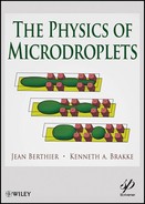
Figure 1 Different applications using microdroplets: (a) droplets moved with magnetic beads for PCR application [6]; (b) protein crystallization in a microdrop (from [7], ©Wiley-VCH Verlag GmbH & Co. KGaA.
Reproduced with permission); (c) encapsulated cells in a polymerized alginate matrix (photo courtesy CEA-LETI); (d) tunable lenses by Varioptics (from [15], courtesy Varioptics); (e) screen displays by Liquavista, (from [16], courtesy Liquavista); (f) schematic of a capillary force actuator (not to scale) [17].

Organization of the Book
This book is dedicated to the study of droplets and interfaces principally in a steady or quasi-steady state, although some dynamic considerations have been added when it was judged useful. The first chapter presents the general considerations leading to the concepts of surface tension and capillary forces, associated to the notions of surface energy and contact angle. Young’s and Laplace’s laws, which are the two “pillars” of any capillary approach, are described, commented and exemplified. The second chapter presents the theory of liquid surfaces in space, including some ways to prove certain surfaces are minimums of energy. Chapter 3 is devoted to the determination of the shape, surface area, and volume of droplets. In chapter 4, the shape and behavior of sessile droplets (droplets place on a solid surface) is investigated for many different configurations of chemical and geometrical surface inhomogeneities: drops at the boundaries between hydrophilic and hydrophobic substrates, or on geometrical inhomogeneities such as steps or grooves or corners. The fifth chapter concerns the behavior of droplets in asymmetric geometries; in a first part, the Hauksbee problem is treated and an extension to hydrophobic surfaces is given. In a second part, the Concus-Finn relations are presented. In chapter 6, the behavior of droplets in microwells and closed microchannels is investigated. The cases of wetting and non-wetting plugs are treated as well as that of trains of droplets. Chapter 7 is dedicated to the phenomena of capillary rise, capillary pumping and capillary valving. In the first two parts, we analyze how capillary forces can contribute to moving a liquid in horizontal or vertical tubes. In the third part, we analyze the opposite: how to find a geometry that can stop a capillary flow. The focus of chapter 8 is open microfluidics, i.e. microflows partially guided by a solid wall, but also in contact with air or another liquid, which is becoming a very important issue in biotechnology; this type of microflow rely mainly on capillary forces and if necessary on electrowetting forces to move the fluid. Chapter 9 deals with the contact and potential engulfment of droplets and particles by interfaces. Examples pertaining to encapsulation of polymerized droplets and capillary assembly are presented. Chapter 10 is on digital microfluidics, a convenient way to manipulate droplets on a planar, or locally planar surface, which has seen many developments lately. We present the state of the art and new developments in this technique. In chapter 11, we treat an example of the use of capillary forces: the ongoing approach to 3D-microelectronics by assembling stacks of chips on a wafer. A promising approach to achieve chip positioning and alignment is that of capillary self-assembly.
References
[1] R. Feynman, Chap 6 in Building biotechnology by Y.E. Friedman, third edition, Logos press, 2008.
[2] P-G de Gennes, F. Brochart-Wyart, D. Quéré. Capillary and wetting phenomena: drops, bubbles, pearls, waves. Springer, 2002.
[3] G.M. Whitesides, Chap 9 in Biotechnology and Materials Science – Chemistry for the future, by L.M. Good, ACS publications, 1988.
[4] P.-A. Auroux, Y. Koc, A. deMello, A. Manz and P. J. R. Day, Miniaturised nucleic acid analysis, Lab Chip 4, pp. 534–546, 2004.
[5] E. Wulff-Burchfield, W.A. Schell, A.E. Eckhardt, M. G. Pollack, Zhishan Hua, J. L. Rouse, V. K. Pamula, Vijay Srinivasan, J. L. Benton, B. D. Alexander, D. A. Wilfret, M. Kraft, C. Cairns, J. R. Perfect, and T. G. Mitchell, “Microfluidic Platform versus Conventional Real-time PCR for the Detection of Mycoplasma pneumoniae in Respiratory Specimens,” Diagnostic microbiology and infectious disease 67(1), pp. 22–29, 2010.
[6] http://www.quantalife.com/technology/ddpcr
[7] Bo Zheng, L. Spencer Roach, and R. F. Ismagilov, “Screening of Protein Crystallization Conditions on a Microfluidic Chip Using Nanoliter-Size Droplets,” JACS 125, pp. 11170–11171, 2003.
[8] T. Thorsen, R. W. Roberts, F. H. Arnold, S. R. Quake, “Dynamic pattern formation in a vesicle-generating microfluidic device,” Phys. Rev. Lett. 86, pp. 4163–4166, 2001.
[9] S.L. Anna, N. Bontoux, and H.A. Stone, “Formation dispersions using flow focusing in microchannels,” Appl. Phys. Lett. 82(3), pp. 364–366, 2003.
[10] J.F. Edd, D. Di Carlo, K.J. Humphry, S. Köster, D. Irimia, D.A. Weitz, M. Toner, “Controlled encapsulation of single cells into monodispersed picoliter drops,” Lab Chip 8(8), pp. 1262–1264, 2008.
[11] D. Witters, N. Vergauwe, S. Vermeir, F. Ceyssens, S. Liekens, R. Puers and J. Lammertyn, “Biofunctionalization of electrowetting-on-dielectric digital microfluidic chips for miniaturized cell-based applications,” Lab Chip 11, pp. 2790–2794, 2011.
[12] H. Song, J. D. Tice, R. F. Ismagilov, “A microfluidic system for controlling reaction networks in time,” Angew. Chem. 42, pp. 767–771, 2003.
[13] A. Gnther, K.F. Jensen, “Multiphase microfluidics: from flow characteristics to chemical and material synthesis,” Lab. Chip 6, pp. 1487–1503, 2006.
[14] J. Atencia, D.J. Beebe, “Controlled microfluidic interfaces,” Nature 437, pp. 648–655, 2005.
[15] VarioticsTM: http://www.varioptic.com/en/tech/technology01.php
[16] LiquavistaTM: http://www.liquavista.com/files/LQV060828XYR-15.pdf
[17] C. R. Knospe and S.A. Nezamoddini, “Capillary force actuation,” J. Micro-Nano Mech. 5 p. 5768, 2009.
[18] O. Lecarme, T. Pinedo-Rivera, K. Berton, J. Berthier, D Peyrade, “Plasmonic coupling in nondipolar gold collidal dimers,” Applied Physics Letters 98, 083122, 2011.
[19] T. Fukushima, T. Tanaka, M. Koyanagi. “3D System Integration Technology and 3D Systems,” Advanced Metallization Conference Proceedings, pp. 479–485, 2009.
[20] K. Sato, T. Seki, S. Hata, A. Shimokohbe, “Self-alignment of microparts using liquid surface tension – behavior of micropart and alignment characteristics,” Precision Engineering 27, pp. 42–50, 2003.
[21] J. Berthier, K. Brakke, F. Grossi, L. Sanchez and L. Di Cioccio, “Self-alignment of silicon chips on wafers: A capillary approach,” JAP 108, 054905, 2010.