Chapter 11
Animal and Plant Poisons
He was a bold man that first ate an oyster.
Jonathan Swift
Massasauga rattlesnake eat brown bread.
Massasauga rattlesnake fall down dead.
If you catch a caterpillar give it apple juice,
But if you catch a rattlesnake, TURN IT LOOSE!
Old Ontario skipping rhyme
Introduction
Chemical warfare is widely practiced in the animal and plant kingdoms. Just as microorganisms produce antibiotics that inhibit the growth and reproduction of competing organisms, more complex plants synthesize chemicals that render them unpalatable to potential predators or that are truly toxic, thus selecting for individuals that avoid them, or producing aversive reactions that limit consumption to once or twice only. Occasionally, however, these plants accidentally enter the food chain of livestock or humans, directly or indirectly, leading to a toxic reaction.
Similarly, toxins and venoms are used in the animal kingdom for defense and prey capture. Toxins are consumed and operate much like those of plants. Toxins can also be secreted by special cells or glands in the skin, so that toxicity may occur simply by taking the intended victim into the mouth. Toxic reactions have occurred among students indulging in the fad of “toad-licking.” These toxins tend to be low molecular weight peptides with neurotoxicity. Some fish actually swim in a “cloud” of toxin secreted by these skin glands. Their toxins are usually steroid glycosides and choline esters. A neurotoxin secreted in this way by a species of flounder of the Red Sea is so powerful that a shark attempting to bite it is incapable of closing its jaws and instantly convulses. It has been proposed as a shark repellant for downed fliers and divers. These skin toxins also serve as antibacterial agents to prevent infection, as the slime secreted by the skin of amphibians is an ideal culture medium for bacteria.
Venoms are injected in some way, and may be employed both for defense and for prey capture. Humans and animals sometimes accidentally become the victims of envenomation or intoxication by poisons of animal origin. The term toxin is used also to refer to the individual components of venoms. Numerous texts have been written on these subjects, and this chapter can do no more than skim the field and discuss some of the more important, or more interesting, examples. Emphasis will be placed on agents that have become important to the biological sciences, either as drugs or as research tools.
Toxic and Venomous Animals
Toxic and Venomous Marine Animals
Venoms and toxins are distinguished by the manner in which they are inflicted upon the victim. A venom is a substance kept in a special poison gland and administered by a complex injection apparatus or by lacerating spines. This type of system may be employed in defense or in prey capture. A toxin is a poison usually ingested as an accidental component of tissues or organs. Toxins likely evolved as a species (rather than an individual) protection. Predators with a preference for that prey would be selected out if the toxin is fatal. Conditioned avoidance could occur in survivors. The term for poisoning by the muscle tissue of scale fish, as opposed to shellfish, is ichthyosarcotoxism. It includes toxins that are accumulated up the food chain from plankton as well as toxins that are synthesized by the fish itself. In the former situation, poisoning is usually by a mixture of toxins.
Scale Fish Toxins
Ciguatoxin
The condition ciguatera, or ciguatera fish poisoning (CFP) is caused by several related toxins that concentrate up the food chain from a photosynthetic dinoflagellate, Gambierdiscus toxicus, to reach toxic levels in large marine fish such as grouper, snapper, amberjack, barracuda, parrot fish, etc. These species are coral reef predators and browsers. CTX neurotoxins are far more potent than the brevetoxins associated with red tides. The LD50 for Ciguatoxin (CTX) is 0.25–4.0 μg/kg versus >100 μg/kg for brevetoxin. Toxic symptoms are gastrointestinal (nausea, vomiting, cramps, diarrhea), neurological (numbness, tingling of lips, throat and tongue, dizziness, headache), and cardiovascular (arrhythmias, cardiac arrest). CTX is thought to increase membrane permeability to sodium and it is responsible for the neurological signs and symptoms. There may be at least 20 different toxins involved in ciguatera fish poisoning. CTX is very toxic to mice that are the test species for detecting its presence. CTXs and brevetoxin are large, colorless, heat-stable molecules classified as ladder-like polyethers, the structure of which has now been elucidated. CTX is 3 nm long. Other unidentified toxins are likely involved in ciguatera poisoning. Ciguatera is by far the most common cause of scale-fish poisoning. An estimated 20,000 people are affected annually in subtropical and tropical climes. Any large, warm water species is a potential source of the toxin.
CTX and brevetoxin bind to a common site on voltage-sensitive sodium channels causing persistent activation and/or prolonged opening of the channel. A component of CTX, maitotoxin (MTX), has recently been purified. MTX is a water-soluble polyether that is a potent inducer of an increase in cytosolic free calcium ion [Ca2+]i in a wide variety of organisms including mammalian cells. MTX appears to activate a voltage-independent, nonselective cation channel that may require an extracellular source of Ca2+ for activation. MTX has become a useful research tool for studying ion channels. MTX is a large, complex, multiringed compound. The LD50 in mice is less than 0.2 μg/kg. This is at least five times more toxic than tetrodotoxin (see also http://www.rehablink.com.ciguatera/poison.htm). Barracuda also sometimes contain a related neurotoxin that is very toxic to cats. In some parts of the West Indies, it is customary to feed some of the meat from a large barracuda to a local cat before humans eat it.
Tetrodotoxin
The term comes from Tetraodontidae, meaning four teeth, the name of the genus that carries the toxin. At least six different neurotoxin receptor sites have been identified on the channel protein for voltage-gated ion channels using the various dinoflagellate neurotoxins. Tetrodotoxin (TTX) toxin is present in puffer fish, which are called “fugu” in Japan. It is a very potent blocker of fast sodium channels, and therefore inhibits nerve conduction in a manner like local anesthetics. It causes paresthesia, paralysis, anesthesia, and loss of speech, but consciousness is retained. Prognosis is improved if vomiting occurs early after ingestion. In Japan there are about 150 cases of poisoning annually with 50% mortality. Fugu is considered a delicacy in Japan and special chefs are licensed to prepare it. The toxin is concentrated in the liver and gonads of the fish. Connoisseurs of fugu prefer that just enough toxin remains to cause a slight tingling of the lips when it is consumed. In Japan, this custom has spawned a somewhat macabre poetry form: “Last night he and I ate fugu; today I helped carry his coffin.”
TTX is also found in some newts and frogs. The blue-ringed octopus, a small octopus that inhabits tidal pools and shallow waters around Australia and other central Indo-Pacific waters, produces a toxin, administered by a bite that is believed to be identical to TTX. The bite of a larger specimen can be fatal in minutes. In recent years, the geographic and species distribution of TTX has widened considerably. Species reported to carry TTX include gobies, gastropods, frogs, starfish, crabs, flatworms, and ribbon worms. Poisoning has occurred not only in Japan but in China, Taiwan, New Zealand, and even Europe. Whether this spread is related to climate change or to alteration of the marine ecology through human activity or is a natural phenomenon is unknown at this time. The toxin is not synthesized by the fish and other marine species but is concentrated up the food chain from marine bacteria. The spread of these bacteria, for whatever reason, could account for the increased number of species affected.
There is a fascinating theory that the zombie tales associated with Haiti arose from poisonings with TTX or from the deliberate of it by voodoo practitioners. Someone who has been poisoned with TTX can appear to be dead but later recover, but in a much impaired state.
TTX is of interest as a research tool because of its potent sodium channel blocking activity. Its occurrence in such diverse species can be explained by the fact that it is not synthesized by the animal itself, but rather by certain species of Vibrio bacteria that exist in a symbiotic relationship with the host.
Scombroid Toxins
The name “scombroid” comes from “Scombridae,” referring to dark-muscled fish like tuna, albacore, and mackerel. The poisoning results from eating fish rich in histidine. However, according to information released by the (U.S.) Centers for Disease control (CDC), the most common vectors are not the Scombridae. Other fish like mahi-mahi and amberjack are frequently responsible. Improper refrigeration results in the decarboxylation of histidine to histamine by surface bacteria. The signs and symptoms resemble a histamine reaction. They may onset in minutes to hours and include dizziness, headache, diarrhea, facial flushing, tachycardia, pruritis, and wheezing. Fish contaminated in this way are often described as having a spicy or peppery taste. Levels of histamine in contaminated fish may exceed 100 mg/100 g. The FDA has set 50 mg/100 g as the hazard level. Histamine is not destroyed by cooking. There is some evidence that another product of decomposition, saurine, may contribute to the symptomatology. Treatment with histamine blockers (H1 and H2) is nearly always successful.
Ichthyotoxin
Ichthyotoxin is a term that is applied to a number of toxins produced by algae and concentrated by scale fish. Freshwater and ocean species are both affected. The chemical nature of many of these toxins remains largely unknown. Karlotoxin-2 is an ichthyotoxin from the dinoflagellate Karlodinium veneficum: It is believed to be responsible for numerous fish kills in an estuarine aquaculture facility in Maryland. During a 1996 fish kill of about 15,000 hybrid striped bass associated with an algal bloom, human health effects were also observed. These included skin lesions, respiratory difficulty, and neurological symptoms primarily consisting of short-term memory loss. Similar past events were generally attributed to the dinoflagellate Pfiesteria piscicida. A highly labile, copper-containing metallo-organic compound has been proposed as the putative P. piscicida toxin but despite countless fish kills in the region a definite link has not been established. The complex chemical nature of karlotoxin-2 has been elucidated. It kills fish in a dose-dependent manner. Karlotoxins were also shown to be cytotoxic and hemolytic.
Red Tide Dinoflagellate Toxicity for Higher Species
Numerous species of dinoflagellate algae can cause the red tides that periodically plague the shores of the Gulf of Mexico and elsewhere. The aerosol created by wave action can cause respiratory problems in people onshore. This can be serious in people with preexisting respiratory problems. Skin contact with the algae can cause an irritating rash. Other animals also can be affected, especially by the aerosol. In the northern Gulf of Mexico, there were 757 dolphin and whale strandings between February 2010 and June 17, 2012. An additional 123 bottle-nosed dolphins washed ashore along the Texas Gulf coast from November 2011 to March 2012. These strandings coincided with a red tide caused by the dinoflagellate Karenia brevis. Although cause and effect has yet to be proven, this temporal association makes the red tide highly suspect as the culprit.
Shellfish Toxins
Dinoflagellates produce a host of toxic substances with a wide array of toxic effects. Their chemical nature is often unknown. The subject of dinoflagellate toxicity was introduced earlier under CTX and Ichthyotoxins. They are also responsible for a variety of shellfish poisonings and the major ones will be reviewed here. An excellent review article by Wang identifies the dinoflagellate responsible for the various toxins. The majority act as neurotoxins.
Saxitoxins
Saxitoxin is the best known of a family of neurotoxins that cause paralytic shellfish poisoning and it concentrates in bivalves such as oysters, clams, and mussels. There have been at least 24 structurally related imidazoline guanidinium derivatives identified that are paralytic shellfish poisons. The mechanism and symptomatology are the same as for tetrodotoxin; they block voltage-gated ion channels (voltage-gated sodium channel 1) for sodium, calcium, and potassium. The LD50 for mice is 3–10 μg/kg intraperitoneally. The lethal oral dose for humans is 1–4 mg. “Never eat oysters in a month without an R” is an old adage that still is good advice in the Northern Hemisphere as dinoflagellates bloom in the summer months.
Brevetoxins
Brevetoxins (there are two main ones) have both gastrointestinal and neurological toxicity and has been responsible for numerous fish kills. These toxins open voltage-gated sodium channels (channel 5) in cell membranes. Signs of poisoning in humans include gastroenteritis, nausea, tingling and numbness around the mouth, loss of motor control, and severe muscle pain.
Domoic Acid
In 1987 there was an outbreak of mussel poisoning in Canada caused by mussels from the waters around Prince Edward Island. The toxin was domoic acid, which concentrates from a seaweed called chondria. It is also found in a diatom and it is rare in North Atlantic waters, being more common in Japan. The toxin was identified by the Canadian National Research Council Atlantic Laboratory. There were three fatalities in elderly nursing home residents in Quebec, and over 100 individuals were affected. Several have been left with short-term memory deficit, a condition called amnesic shellfish poisoning. It has been suggested that marine pollution might have changed the environment to favor the growth of the offending diatom. Domoic acid binds to a specific subset of glutamic acid receptors, known as kainate receptors, in the brain.
Okadaic Acid
Also produced by a dinoflagellate, okadaic acid is responsible for a condition known as diarrhetic shellfish poisoning. There is some evidence that there has been a dramatic increase in toxic algal blooms, the so-called red tides. The first confirmed outbreak of diarrhetic shellfish poisoning in North America occurred in 1990 and it was traced to dinoflagellates in Canadian waters. Brown pelicans eating anchovies off California were dying of domoic acid poisoning in 1991, saxitoxin has been found in Alaska crabs, and in 1987–1988 shell fishing off North Carolina was shut down because of a red tide of a dinoflagellate that produces a neurotoxin, brevetoxin (see also under CTX in the preceding text).
Okadaic acid is a selective inhibitor of phosphatases 1 and 2A with interference to protein activation/inactivation. It can induce contraction of smooth muscle and cardiac muscle. It has been shown experimentally to be a tumor promoter but, unlike phorbol esters, it does not activate protein kinase C.
Azaspiracid Toxin
Shellfish poisoning with Azaspiracid toxin (AZP) toxin was first reported in the Netherlands from eating Irish mussels in the 1990s but has since become a widespread problem throughout Europe. It is produced by the dinoflagellate Protoperidinium crassipes. There have been over a dozen derivatives of this unique lipophilic, polyether toxin. Symptoms of poisoning are primarily gastrointestinal (nausea, vomiting, severe diarrhea, stomach cramps) but neurological symptoms may also occur. Experiments in mice have shown that repeated injections may cause the formation of lung tumors. The mechanism of action is presently unknown.
Yessotoxin
Yessotoxin (YTX) and its analogues are disulfated polyethers and are becoming more commonly associated with shellfish poisoning, having been identified in Japan, New Zealand, Europe, and South America. They were originally thought to be responsible for the diarrheic shellfish poisoning (DSP) because they were isolated along with other DSP toxins. Animal studies did not confirm this effect but indicated that the heart was the target organ. Neurotoxicity has also been shown experimentally.
Palytoxin
Palytoxin (PTX) is a very large, highly complex polycyclic compound that has biological activity at extremely low concentrations. It is believed to have the longest continuous chain of carbon atoms of any natural substance. Originally isolated from a soft coral, it has since been identified in seaweeds, shellfish, and other marine organisms including a benthic dinoflagellate, Ostreopsis siamensis, which causes algal blooms along the coast of Europe. It is of increasing concern for both economic and public health reasons. It has caused excessive die-offs of edible mollusks and echinoderms, as well as illness in people. Deaths from PTX have been recorded from eating contaminated crabs in the Philippines, sea urchins in Brazil, and fish in Japan. The mechanism of action is unknown but symptoms include weakness, ataxia, drowsiness, fever, and death. The intravenous LD50 in experimental laboratory animals ranges from 0.25 to 0.9 μg/kg.
Stinging Fish Venoms
In venomous fish such as the stonefish, lionfish, scorpionfish and stingray, spines are located ahead of the dorsal fin, on the tail, or around the mouth. A heat-labile protein causes intense pain and cardiac shock. Heat above 50°C may afford some relief. Stonefish and stingrays are most often stepped upon because they conceal themselves in the sand bottom. Fatalities have occurred from envenomation by these fish, generally as a result of cardiovascular shock, with AV block and bradycardia. Other symptoms include numbness, inflammation and edema at the site of injury, severe pain in surrounding tissues, delirium, nausea, vomiting, and sweating. Stingray venom is a large, heat-labile protein (molecular weight >100,000) with neurotoxic as well as cardiotoxic properties. There is an antivenin for stonefish venom. Figure 11.1 shows a scorpion fish, one of the less hazardous members of this group.
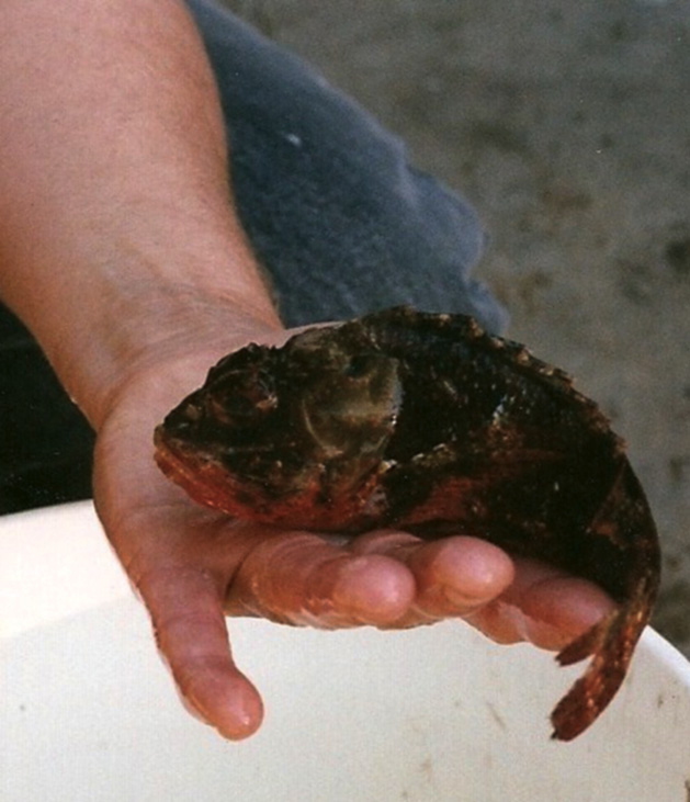
A staff member of the Gulf Specimen Marine Laboratory holds a scorpion fish in the palm of her hand. The dorsal spines (folded back here) are the means of conveying the venom into tissues.
Mollusk Venoms
Conotoxins
These are found in marine cone snails. All are strongly basic peptides highly cross-linked by disulfide bonds. Alpha-conotoxins are nondepolarizing, neuromuscular blocking agents like curare, and they therefore cause paralysis. They are 13–15 amino acid peptides. Mu-conotoxins are 22 amino acid peptides that block sodium channels, thus acting like tetrodotoxin and saxitoxin. These sodium channel blocking conopeptides are under investigation for use in treating chronic pain. Omega-conotoxins block presynaptic, voltage-dependent calcium channels. Recent research has identified a number of subtypes with specificity for various channel subtypes, making them useful research tools. Cone snails are univalve gastropods with a complex envenomation apparatus they use for prey capture (see http://grimwade.biochem.unimelb.edu.au/cone/newlog.html).
Cone shells such as the cloth of gold cone shell are highly prized by collectors. The envenomation apparatus can reach any exterior point on the shell and collectors have been seriously envenomated and even killed by handling live cone shells of the most toxic types.
Coelenterate Toxins
Many coelenterates (anemones, sea urchins, jellyfish, and corals) produce venoms that can cause pain on contact or even systemic envenomation. Fire coral produces a protein venom that causes intense local burning when touched. It feels like a cigarette burn (as this author can attest personally).
Physalia physalis (Portuguese man-o’-war, bluebottle, mauve stinger) is not a true jellyfish but rather belongs to the siphonophores, which are not single organisms but colonial ones composed of many single-celled units called zooids. These are linked together and physiologically interdependent. The bluebottle drifts on the wind by virtue of its sail which protrudes above the surface of the water. The bluebottle causes signs and symptoms like fire coral. Red streaks, called straps, occur where tentacles touch skin. Allergic reactions can occur, also generalized symptoms such as fever, nausea, and cardiac and respiratory distress. Bluebottles washed up on beaches remain toxic until completely dried out. First aid consists of washing the affected area with salt water followed by hot water (45°C, 115°F) both of which denature the venom. Even urine may help relieve the pain. This species is common around the world. Vinegar is not recommended as it promotes further release of venom from the nematocysts.
True jellyfish belong to the phylum Cnidaria. They are free-swimming animals with an umbrella-like bell that pulses to provide a form of jet propulsion. Although lacking a brain or true nervous system, jellyfish do have a loose nerve net and appear able to sense some environmental conditions. The sea wasp (Chironex fleckeri, box jellyfish) is the most venomous marine animal known. It inhabits the Indo-Pacific region and accounts for many deaths annually. In Australia, a registry has been kept since the mid-twentieth century and about 70 deaths have been attributed to the sea wasp. Contact with tentacles causes intense, agonizing pain, coma, and cardiac shock. Mortality is 25% and children, the elderly, and heart patients are most vulnerable. First aid is denaturation with vinegar, removal of tentacles using forceps, gloves, a knife and fork, or any means to avoid touching the tentacles, and cardiopulmonary resuscitation (CPR) if required. CPR should be continued for as long as possible as late recovery has been reported. A specific antivenom for the sea wasp is available. Local anesthetic spray such as is used for sunburn may be helpful.
The “Irukandji” (Carukia barnesi and Malo kingi) is a tiny, four-tentacled, Indo-Pacific jellyfish that causes similar signs and symptoms as the sea wasp plus massive sympathetic discharge. Irukandji syndrome is characterized by a massive release of adrenaline (epinephrine) and other catecholamines and histamine. This jellyfish can actually inject barbs into the skin and cause systemic symptoms including hypertension. First aid consists of vinegar washing, which denatures the venom on the surface but not that already injected. Antihistamines may be helpful.
Echinoderm Venoms
Sea urchins and sea anemones such as the long-spined or black sea urchin, and crown-of-thorns sea urchin possess toxins. Injury usually occurs to the feet and lower limbs when a diver or swimmer steps on these bottom dwellers. Spines are driven into the flesh and break off. The toxin produces local pain and burning similar to that of the bluebottle. It is heat labile, so immersing the affected part in water as hot as the person can stand may help. Unlike the usual first aid for envenomations, movement and trauma may actually help by breaking up the spines so they can be absorbed more quickly. Vinegar or even urine may also help.
Sea anemones also produce very potent toxins that act when they are ingested. Best characterized of these are the equinatoxins (EqTs I, II, and III) from Actinia equina. In rats they cause coronary vasospasm, cardiac arrest, and other cardiorespiratory toxicity. Hemolysis also occurs, and degranulation of blood platelets and white cells.
For more information on marine venoms and toxins see http://www.pmeh.uiowa.edu/fuortes/63260/MARINEAN/index.htm and http://www.merck.com/pubs/mmanual/
Freshwater Algae
Cyanobacteria comprise a group of prokaryotes that share characteristics of both bacteria and algae, leading to a plethora of often confusing names. Here they shall be referred to by their most familiar name, the blue-green algae. Like our own species, these organisms are capable of being highly beneficial as well as causing harm. They are important primary producers with high nutritive value. Nitrogen-fixing species contribute to both soil and water fertility. Future use in food production and solar energy conversion has been proposed to be an important, future application.
On the opposite side of the coin, they can contaminate water reservoirs and pose a threat to drinking water supplies and human health. Records from the Han dynasty in China (c. 950) noted the death of troops who drank from a green river. Reports of poisoning of domestic animals are numerous. Eutrophication of water bodies usually related to high phosphate levels results in algal blooms and scum formation. Discharges from municipal wastewater discharges and agricultural runoff accelerate the process. At least 85 toxins from this group have been identified. Hepatotoxins are dominant (microcystins and nodularins). They are protein phosphatases and tumor promoters. Neurotoxins have also been identified. These include mimickers of acetylcholine, an anticholinesterase and even saxitoxin, the toxin of paralytic shellfish poisoning (see later).
Fish kills from cyanobacteria are common. A recent local kill occurred in a small stream killing hundreds of fish of several common freshwater species including rock bass, chub, and white suckers.
Prymnesium parvum (golden algae) has spread extensively to inland waterways in southern North America and has been associated with fish kill events. It is migrating northward and has been responsible for ecological devastation that threatens the economic and recreational value of freshwater systems. Studies have shown that the array of toxins produced by cultured P. parvum is markedly different from that produced by wild organisms. A highly labile Ichthyotoxin was present in both, however, and was proposed by the authors as the probable cause of fish kills.
As noted earlier, algae can be highly beneficial as well as harmful. A unique example of this came to light recently. There is a very large cement plant in the area. It is now directing its smokestack emissions to a 20,000 L tank on site. Algae, whose growth is stimulated with a specific wavelength of light, utilize the carbon dioxide, nitrogen oxide, and sulfur dioxide for growth. The biomass is compressed to extrude an oil that can be used as biodiesel and the residue is dried and made into bricks that burn like coal. Cement plants are believed to cause about 5% of global carbon dioxide emissions. When the facility is at full production it will produce about 225,000 ton of biomass annually.
Toxic and Venomous Land Animals
Venomous Snakes
Fear of snakes (ophidiophobia) is a very common phobia. Some psychologists feel it has its origins in the biblical story of the Garden of Eden, but the fear may not be all that irrational. It is estimated that 50–100 thousand people die annually from snakebite and thousands more endure permanent disability. Tropical and subtropical areas record the most bites and rural dwellers are most often affected. Farmers working in their fields, often barefoot, are especially vulnerable. Many snakes are venomous, but most have rear fangs that are designed to paralyze prey after it has been taken into the oral cavity. These are incapable of inflicting a venomous bite (Figure 11.2). The four genuses of poisonous snakes that are a danger to humans are Viperidae, Crotalidae, Elapidae, and Hydrophiidae.
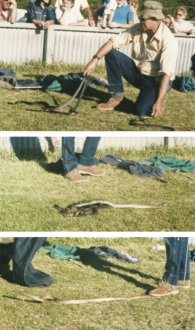
Top: An Australian snake handler is holding three, red-bellied black snakes (Pseudoechis porphyriacus ). While venomous, the bite of this snake is rarely fatal and it is not too aggressive. Middle: Two tiger snakes (Notechis sp.) have been just released from their bag by the handler. This snake is highly venomous and the mortality if untreated with anti-venom is 40 to 60%. Bottom: The handler restrains this eastern brown snake (Pseudonaja sp.), considered to be the second most venomous land snake. It can grow to two meters and is very fast. Mortality is similar to the tiger snake. All of these snakes are elapids.
- Viperidae. These Old World vipers have hollow, needle-like fangs that are set in short, movable maxilla that rotate to bring the fangs into the biting position as the mouth is opened. The head is large and triangular. Signs and symptoms of the bite are as for the Crotalidae. Viper bites are not uncommon in Europe, occurring most often in children. One French paper reported on 58 children bitten by adders (vipers) between 2001 and 2009. Moderate to severe envenomation occurred in 17% of the patients. Bites to the upper extremities were associated with more severe envenomation. A polyvalent antivenin, Viperfav (Vipera aspis, Vipera berus, Vipera ammodytes), was used in severe cases.
- Crotalidae (or Crotalinae). The Peterson Field Guide on North American Venomous Animals and Poisonous Plants lists 31 species and subspecies of rattlesnakes, three of cottonmouths, and five of copperheads. These “pit vipers” are similar to the Old World vipers with folding fangs but they also have a deep, infrared-sensitive pit between the eye and the nostril that is used for tracking prey by body heat. This genus includes all rattlesnakes, the water moccasin (or cottonmouth) and copperhead. All are found in North America. Canada has the Western Diamondback and, in Ontario, the Massasauga rattlesnake. The timber rattlesnake is thought to be extinct but some feel it may have survived in areas of the Niagara Escarpment. The United States has about 25 species of venomous snakes and most are crotalids. About 8000 venomous snakebites occur annually with several deaths.
Signs and symptoms of envenomation include tissue swelling at the site, pain, ecchymosis (purplish, streaky hemorrhages into the skin), altered mental status, tachycardia, respiratory distress, and hypotension. Laboratory tests show evidence of coagulopathy (hypofibrinogenemia, low platelet count, prolonged clotting tests, e.g., activated partial thromboplastin time). A polyvalent antivenin now is available, FabAV or Crofab. It is composed of the Fab fragment of antibodies derived from ovine sources immunized with venoms from the Western Diamondback rattlesnake (Crotalus atrox), the Eastern Diamondback (Crotalus adamanteus), the Mojave rattlesnake (Crotalus scutulatus), and the Cottonmouth (Agkistrodon piscivorus). FabAV has much lower allergenic potential than older, polyvalent crotalid antivenin. Coagulopathy following pit viper envenomation may persist or recur for up to 2 weeks and require periodic treatment with Fab-based antivenins.
Pit vipers can be found worldwide. Asian species include the Chinese pit viper (Protobothrops mangshanensis) and the Green pit viper (Trimeresurus albolabris, Trimeresurus trigonocephalus). In Mali, a study in two national hospitals recorded 832 snakebites from 1993 to 2002. The offending species were not identified but the symptomatology was characteristic of viper venom. Through the use of antivenom the mortality rate was reduced from 12% in 1995 to 3% in 2002.
- Elapidae. They are distributed worldwide and account for the most venomous and feared snakes of the tropics and subtropics. This group includes the cobras, the boomslang, and many Australian species (taipan, tiger snake, brown snake, etc.). There are three species in the United States: the eastern or Florida coral snake (Micrurus fulvius), the Arizona coral snake (Micruroides euryxanthus), and the Texas coral snake (Micrurus tener) that apparently is found only in Harris County. The rhyme “red-on-black, friend of Jack, red-on-yellow, kills a fellow” helps to distinguish the coral snake from harmless look-alikes, such as the scarlet king snake and other king snakes and the Louisiana milk snake. This rhyme, however, applies only to North American coral snakes. The markings of the South American ones do not always have the typical pattern. Coral snakes have hollow, short, rear fangs and tend to hang on rather than strike and release like vipers. Bites are rare.
- Hydrophiidae (or Hydrophiinae). There are some 50 species of sea snakes distributed worldwide. They are restricted to the warmer waters of the Pacific and Indian Oceans. Most are marine dwellers but some may enter river estuaries. All but one genus (Laticauda, which lays eggs onshore) give birth to live young in the water (ovoviviparous). Although they are highly venomous, they are not usually aggressive except during mating season. Most bites occur to commercial fishermen because the snakes become trapped in the nets or when the fishers are wading in muddy waters. For some years, sporadic discussions have occurred concerning the feasibility of digging a sea-level canal across the Isthmus of Panama to connect the Atlantic and Pacific Oceans. One environmental concern that has been raised is that such a canal could introduce the yellow-bellied sea snake (Pelamis platurus) to the Gulf of Mexico and the Caribbean Ocean where conditions are favorable for their proliferation. This sea snake may occur inshore from southern California through Central and northern South America. Claims of sea snake sightings around Jamaica are thought to be due to mistaking the spotted eel for a snake. The clinical picture of sea snake envenomation includes rhabdomyolysis (dissolution of muscle tissue) with resulting myoglobinemia and myoglobinuria, muscle pain and tenderness, flaccid paralysis (neurotoxicity), and renal failure. Antivenom is available.
- Trimorpohodon spp. In addition to the coral snakes there are other, venomous, rear-fanged snakes that rarely cause envenomation as they have difficulty reaching human limbs with their fangs. These so-called lyre snakes, so named from a lyre-shaped marking on the back of the head, include the Texas lyre snake, the Sonoran lyre snake, and the California lyre snake. Small children could conceivably be bitten on the finger due to their small size but this author is unaware of any recorded incidents.
- Heloderma spp. Gila monsters are native to the desert areas of the American southwest and Mexico. There are two subspecies, Heloderma suspectum suspectum (reticulate) and Heloderma suspectum cinctum (banded). Venom glands are located at the base of the grooved teeth and Gilas have a tendency to hang on and “chew in” the venom. Gilas are not generally as dangerous as venomous snakes. The venom lacks neurotoxins but contains coagulants and enzymes as well as serotonin. The reaction tends to be more local. Lethal doses in animals lead to cardiorespiratory collapse.
Snake Venoms
Over the last decade a massive literature has developed concerning research into the chemical nature and biological activities of snake venoms. Given the very large numbers of these, it is only possible to skim the surface of the field here. Snake venoms are complex mixtures of proteins and polypeptides, many of which are proteolytic enzymes. In general, venoms of the Elapidae and the Hydrophiidae tend to be neurotoxic with myonecrosis (breakdown of muscle tissue) occurring at the bite wound, whereas venoms of the vipers and crotalids are generalized coagulants with local anticoagulant activity to spread the venom, causing much local damage (pain, necrosis, bleeding). Neurotoxicity is less prominent.
Signs and symptoms of neurotoxic venoms include progressive paralysis, muscle spasm, respiratory distress or failure, muscle ache (myalgia), kidney failure with myoglobinuria (the appearance of myoglobin in the urine), and cardiac failure. These symptoms apply to coral snakebites in North America. The venom of the Banded Krait (Bungarus fasciatus), native to the Indian subcontinent, contains alpha-bungarotoxin, which is an irreversible blocker of acetylcholine receptors, and beta-bungarotoxin that causes a massive release of neurotransmitter vesicles. Alpha-bungarotoxin is used as a research tool in biomedical research. The Black Mamba (Dendroaspis polylepis) produces dendrotoxin-I, a K+ channel blocker.
Signs and symptoms of viper and crotalid bites (including the Massasauga rattlesnake) include immediate, intense burning at the site of envenomation (like a bee sting), followed by numbness, swelling, shock, and hematuria. Necrosis and possibly gangrene may occur later at the bite wound. Subcutaneous hemorrhages may be present and severe cases may show signs of neurotoxicity. Table 11.1 lists some of the major components that have been identified in snake venoms. The venom of Russell’s viper contains an activator of clotting Factor X and it is used in coagulation research.
Some Components of Snake Venoms
|
Component |
Elapidae/Hydrophiidae |
Viperidae/Crotalinae |
|
Nicotine blocker (neuromuscular blockade) |
+ |
+/– |
|
Cholinesterase (neurotoxic) |
+ |
– |
|
Coagulant protease (increases clotting) |
– |
+ |
|
Anticoagulant protease (decreases local clotting) |
+/– |
+ |
|
Adenosine triphosphate (ATP) (shock, hemolysis) |
+ |
+ |
|
Phospholipase (shock, hemolysis) |
+ |
+ |
|
Phospholipase A (release of histamine and leukotrienes) |
+ |
– |
|
Bradykininogen (forms bradykinin, causes pain) |
– |
+ |
|
Hyaluronidase (breaks down interstitial glue, spreads venom) |
+ |
+ |
Phospholipase A2 complexes in rattlesnake venoms are neurotoxic as well as induce tissue damage. Myotoxic components have exhibited mitogenic multiple effects on growing cultured myocytes. Two potent beta-neurotoxins, crotoxin (from Crotalus durissus terrificus) and ammodytoxin (from V. ammodytes) were recently characterized by x-ray crystallography and their three-dimensional structure determined. The active molecular sites for both neurotoxic and anticoagulant activities were identified.
Elapidae and Viperidae both possess α-neurotoxins that act postsynaptically to prevent acetylcholine from binding to its receptor, as well as neurotoxins that affect transmitter release presynaptically; beta-neurotoxins have phospholipase A2 activity. The mambas and other African snakes have voltage-dependent K+ channel blockers (dendrotoxins), noncompetitive inhibitors of acetylcholine (fasciculins), muscarinic toxins, and L-type Ca++ channel blockers (caliseptins). All are small proteins containing about 60 amino acids and three or four sulfides.
Neurotoxins from venom of the sea snake Laticauda semifasciata, alpha- and beta-erabutoxin have been shown to belong to a superfamily of long-chain and short-chain amino acid neurotoxins and cytotoxins found in both sea snakes and terrestrial snakes. The erbutoxins are short amino acid chain ones that block postsynaptic, nicotinic acetylcholine receptors like curare.
As the chemical structures of snake venoms are slowly unraveled, it is hoped that this may lead to further advances in treatment and possibly even prevention. New tools for pharmacological research may also emerge and possibly new treatments for clotting disorders.
Table 11.1 lists some major components of snake venoms.
First Aid
Regardless of the type of snakebite, the most important first aid measure is a tension bandage applied to the entire affected limb with the same tension one would use to bandage a sprain. Splint immobilization may also be helpful if practical. Rest and reassurance are important. Transportation to a medical center possessing antivenin should occur as soon as possible. Modern hospitals should have the antivenin or polyvalent antivenin, appropriate to their area, in stock. Forced exercise and alcoholic beverages are definitely contraindicated, as is incision and suction at the site of envenomation.
Venomous Arthropods
Members of the order Hymenoptera (bees, wasps, ants) of the class Insecta, and of the class Arachnida (spiders, scorpions) have venoms that contain substances commonly involved in the mammalian pain response. The Old World scorpion Leiurus quinquestriatus produces charybdotoxin that affects calcium-activated K+ channels as does the Israeli yellow scorpion (Leiurus q. hebraeus), and the bee venom apamin. The Mexican scorpion Centroides noxius produces noxiustoxin, which blocks voltage-dependent K+ channels (see Table 11.2). Mast cell degranulating peptide in bee venom also blocks these channels. Scorpion toxins that can target voltage-gated sodium channels consist of 60–76 amino acids cross-linked by four disulfide bridges. They constitute two groups classed as alpha and beta toxins. A very venomous Brazilian scorpion (Tityus serrulatus) contains a beta toxin, Ts2.
Components of Hymenoptera Venoms
|
Hymenoptera |
Histamine Releaser |
Bradykininogen/Bradykinin |
Serotonin |
K+ Channel Blocker |
Melittin, Hyaluronidase, PLA2 |
|
Bees |
Apamin (Ca+ activated) Mast cell degranulating peptide (voltage-activated) |
+ |
|||
|
Ants |
+ |
+ |
+ |
||
|
Wasps |
+ |
+ |
+ |
||
|
Old world scorpions |
+ |
+ |
+ |
Charybdotoxin (Ca+ activated) R-agitoxin-2 (voltage-activated) |
Voltage-activated Na + Channels + |
|
Mexican scorpions |
Noxiustoxin (voltage-activated) |
+ |
Allergic reactions may occur to any of these insect venoms and may be life threatening if severe or if multiple stings occur or if the sting is in the throat. Direct toxic effects account for less than 5% of all deaths from hymenopteran stings. The African honeybee is not dangerous because it is more venomous but because it is extremely aggressive so that multiple stings are common. In North America about 3.3% of adults and 0.8% of children are allergic to honeybee and wasp venom. Signs and symptoms include flushing, tachycardia, abdominal colic, diarrhea, and, in severe cases, progressing to hypotension and coma. Severe cases require injection of epinephrine (adrenaline) with an Epi-pen, antihistamines, and corticosteroids. There have been about 20 identified enzymes, peptides, and active amines identified in bee venom. These include phospholipase A2 and hyaluronidase, which breaks down hyaluronic acid, the intercellular glue that maintains conformational integrity. Hyaluronidase acts as a spreading agent. Phospholipase A2 disrupts cell membranes. Mass bee attacks result in the release of large amounts of cytokines like interleukins 1, 6, and 8, and tumor necrosis factor (TNF).
All spiders are venomous but most lack jaws powerful enough to penetrate human skin or do not inject enough venom to cause anything more than some local irritation. Neurotoxins tend to predominate in spider venoms. So-called widow spiders (Latrodectus spp.) have a worldwide distribution in temperate and tropical climes. The black widow, also known as the death spider, hourglass spider, and by many other names, inhabits all of Ontario south of Sudbury but it is rarely encountered since the demise of the outdoor privy. The venom is extremely toxic but very little is injected so fatalities are rare. The toxin is complex. At least seven “latrotoxins” have been identified; five are specific for insects, one for crustaceans, and one for vertebrates. The latter is alpha-latrotoxin (α-LTX). It is a medium-sized protein now available in pure form (see later) that induces the release of most if not all neurotransmitters. It bears similarity to beta-bungarotoxin (found in the Banded Krait). Both cause a massive release of cholinergic vesicles.
Fasciculations and board-like rigidity of the muscles of the trunk occur rapidly followed by muscle cramps, pain in the muscles, and respiratory distress. The presence of a specific receptor on vertebrate neurons for α-LTX has been identified and this is the likely explanation for the species specificity of the toxin. Recovery from black widow spider bite takes about 12 h.
The brown recluse (Loxosceles spp.) known also as the violin spider, has been working its way north from Mexico and Florida and has reached New York State and probably southern Ontario aided, no doubt, by climate change. The bite is painless. Local necrosis develops and expands over the next week due to local blood clotting and microthrombosis. Permanent scarring may result. Occasional deaths have occurred from hemolytic anemia and kidney failure. The venom is complex and includes coagulants, enzymes, and a complement inhibitor.
Funnel web spiders (Atrax spp. and Hadronyche spp.) from southeast Australia give a bite that resembles a scorpion sting and is characterized by massive catecholamine release. Banana spiders (Phoneutria nigriventer) can be shipped in banana bunches and cause serious bites, especially in countries where no immunity is likely. Funnel web and red-backed spiders are claimed to be especially venomous. Two funnel web toxins have been identified, a polyamine (FTX) and omega-agitoxin. Both block high-voltage-dependent P Ca2+ channels, and they are now used as research tools for this purpose. R-agitoxin-2, from the Israeli yellow scorpion, blocks some voltage-activated K+ channels.
Toxic Plants and Mushrooms
Introduction
Folk knowledge, passed orally from generation to generation, usually determines what we can eat and what we cannot eat, and even what parts of a plant are edible. Thus, we make pies from rhubarb stalks but we never make salad from the leaves, which are toxic. Nor do we eat the bulbs or stalks of the many ornamental flowers and shrubs in our gardens. Seldom do we consider the fact that these decisions are toxicologically based. The number of plants that are potentially harmful is too voluminous for extensive coverage. The subject has formed the basis of many texts. The Peterson field guide, Venomous Animals and Poisonous Plants by Foster and Caras lists over 250 species of plants and mushrooms with potential toxicity and concedes that it is by no means exhaustive. The agricultural costs from livestock consuming toxic plants can be extreme. One study reported that in the 17 southwestern states of the United States, direct losses were estimated at $340 million annually using 1989 data. This figure did not include indirect losses due to such things as poor growth rate, prolonged gestation period, infertility, etc. nor costs of preventing poisoning through fencing, destruction of weeds, and other measures.
The following list contains examples of major groups of toxicants chosen because they are common or because of their pharmacological significance.
Vesicants
Many plants contain oxalates that can cause corrosive burns to the mouth, esophagus, and stomach. Symptoms also include vomiting and diarrhea. Since oxalates are anticoagulant, bleeding may also occur. Dieffenbachia contains calcium oxalate, which is a vesicant. Rhubarb leaves contain a variety of oxalates and may be fatal if consumed in quantity due to their renal toxicity (see also ethylene glycol). May apple, buttercup, and philodendron contain vesicants. Philodendron sometimes causes poisoning in cats if they chew the leaves. Poison ivy, poison oak, and poison sumac all contain urushiol, which is a phenolic vesicant.
Cardiac Glycosides
White and purple foxgloves (Digitalis lanata and D. purpura) are the commercial source of medicinal digitalis. Many garden plants possess similar active components, including lily of the valley, star of Bethlehem, and oleander. Symptoms are those of digitalis overdose; nausea, vomiting, visual disturbances (a green halo seen around objects), and cardiac arrhythmias.
Astringents and Gastrointestinal Irritants (Pyrogallol Tannins)
Acorns, geraniums, sumac berries, hemlock bark, rhubarb leaves, horse chestnut, all contain these. North American natives learned to remove tannins from acorns by steeping the crushed nuts repeatedly in freshwater. This will not work for horse chestnuts, however.
Autonomic Agents
Deadly and wooded nightshades contain atropine (hyoscyamine) and scopolamine (hyoscine). The signs and symptoms are those of blockade of muscarinic, cholinergic receptors plus CNS symptoms (disorientation, hallucinations). Poisoning from consuming the berries continues to occur, usually in children. A group from Turkey reported on 49 poisoned children, six of whom were considered to be severely affected. Signs and symptoms included meaningless speech, tachycardia, mydriasis, flushing, and in severe cases, coma. Artificial ventilation was not necessary for any patient and all recovered fully. As physostigmine was not available, neostigmine was used successfully. Practitioners of folk medicine sometimes use belladonna with potentially harmful consequences. A report from Africa (Morocco) deals with an 11-year-old girl under treatment for tuberculosis (rifampicin and isoniazid). She was given a preparation of Atropa belladonna by a herbalist. She presented at the local hospital 24 h later with dry mouth, incoherent speech, confusion, inability to recognize members of her family, uncontrollable vomiting, visual hallucinations, visual and hearing disturbances, and intermittent coma. She was treated successfully with symptomatic treatment that included oxygen, diazepam (tranquilizer/sedative), antiemetics, and i.v. electrolytes.
If ingestion has been recent, activated charcoal is often given orally to neutralize any toxin still in the gastrointestinal tract.
Amanita muscaria is the common poisonous mushroom. Signs of poisoning are those of massive cholinergic discharge due to the toxin muscarine, plus CNS effects like those of nightshade due to the presence of anticholinergic agents. CNS depression and cardiac failure may follow. Symptoms may be delayed 12–24 h.
Solanine and chaconine are glycogenic alkaloids that occur in potatoes and tomatoes (members of the Solanaceae family) when they are exposed to excessive sunlight, blight, sprouting, and prolonged storage. The compounds have cholinergic activity and have been shown to be teratogenic. Symptoms of poisoning include vomiting, diarrhea, cramps, dizziness, visual disturbances, and other CNS manifestations. In 1820, a man named Johnson defied conventional wisdom by publicly eating a tomato in Salem, New Jersey. To the surprise of onlookers, he did not die a horrible death. At the time it was widely believed that tomatoes, because of their relationship to deadly nightshade, were highly toxic. Johnson single-handedly launched the North American tomato industry.
Another member of the Solanaceae is Datura stramonium, commonly known as jimsonweed or thorn apple. Other names are devil’s trumpet and angel’s trumpet. This is a common weed throughout North and Central America, although it was first introduced from Europe. It typically grows in rough ground such as vacant lots. It has a large, white or mauve trumpet flower and a spiny seedpod. The seeds can contain significant levels of atropine and scopolamine that can induce hallucinations. For this reason it is sometimes used as a recreational drug, usually by adolescents. Due to its anticholinergic activity, signs and symptoms of jimsonweed poisoning can include dilated pupils, dry mouth, rapid and irregular heartbeat, stasis of the bowel, agitation and disorientation, as well as hallucinations. Several deaths have occurred.
Dissolvers of Microtubles
Colchicine from the autumn crocus is used in the treatment of gouty arthritis and as an experimental tool. It dissolves microtubules to prevent mitosis and also phagocytosis. Overdose causes severe diarrhea. Vincristine and vinblastine from the periwinkle plant share this property and are used to treat childhood leukemias. Podophyllotoxin from the May apple also dissolves microtubules.
Phorbol Esters (for Example, Phorbol Myristate Acetate, PMA)
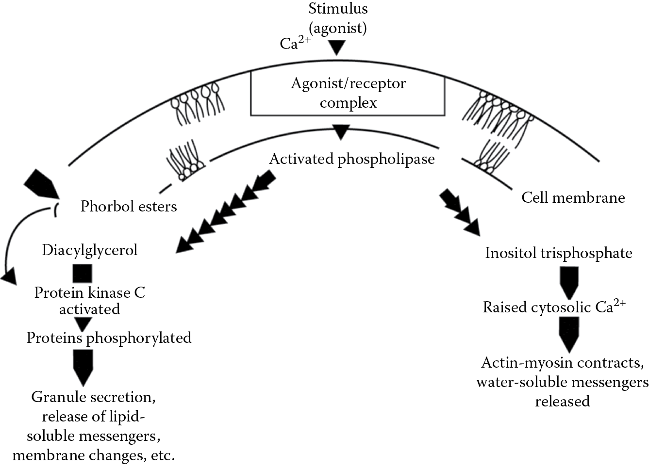
These are components of croton oil from spurge plants. These substances are cancer promoters. They directly activate protein kinase C (substituting for diacylglycerol) independently of extracellular calcium and are they are used as experimental tools for this reason. They act as drastic purgatives. The site of action of PMA is shown in Figure 11.3. It affects an important control mechanism for intracellular regulation.
Many other carcinogens, co-carcinogens, promoters, and anticarcinogens exist in plants. Safrol is a liver carcinogen found in some spices (nutmeg, cinnamon) and in oil of anise (licorice flavoring). The use of anise oil and of oil of sassafras has been banned. Some tannins are liver carcinogens, and the polyaromatic hydrocarbon (PAH) benzo-[a]-pyrene is a potent carcinogen that occurs in green vegetables, unrefined vegetable oils, coconut oil, and chicory. Benzanthracenes are other PAHs that occur in vegetables. Many others exist.
Thapsigargin is a plant-derived sesquiterpene lactone capable of inhibiting Ca2+-ATPase and causing discharge of internal Ca2+ stores. It is widely used as a research tool because of this action. It also has tumor-promoting properties.
Cyanogenic Glycosides
Substances such as amygdalin in almonds, dhurrin in sorghum, linamarin and lotaustralin in cassava and lima beans, and prunasin in stone fruit (cherries, peaches, and chokecherries) are cyanogenic glycosides. They are capable of forming hydrogen cyanide (HCN) with the beta-glucuronidases from the plants when cells break down or from the microflora of the gastrointestinal tract. Cyanide poisoning can occur in ruminant animals from eating vegetation high in cyanogenic glycosides or in humans who have consumed improperly stored or prepared foods such as lima beans or cassava. In humans, CN can be formed from organic nitriles by Cytochrome P450-dependent mono-oxygenases, and from organic thiocyanates by glutathione S-transferases.
Detoxification of Hydrogen Cyanide
Hydrogen cyanide (HCN) is detoxified by conversion to thiocyanate, which requires sulfur-containing amino acids and vitamin B12. Deficiencies of these increase the risk of toxicity. The metabolic detoxification system is overwhelmed and hydrogen cyanide interferes with electron transport in the cytochrome a–a3 complex, with resulting in tissue hypoxia. This leads to rapid failure of the CNS and death. Treatment is the administration of intravenous nitrites, which form methemoglobin. Methemoglobin has a high affinity for HCN and binds it to protect the cytochrome and allow time for biotransformation to occur. These events are summarized in Figure 11.4.
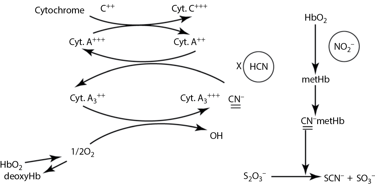
Site of action of HCN and of detoxification by nitrites.
Used in Research and Treatment
Many chemicals of animal and plant origin are useful as research tools in physiology and pharmacology. A partial list follows.
- Tetrodotoxin from puffer fish and saxitoxin from shellfish block fast sodium channels and are used to study nerve conduction.
- Alpha-conotoxin from cone snails is a nondepolarizing neuromuscular blocking agent.
- Mu-conotoxin from cone snails acts like tetrodotoxin.
- Omega-conotoxin from cone snails is a specific inhibitor of presynaptic, voltage-dependent Ca2+ channels.
- Russell’s viper venom activates Factor X in the clotting system and is used in certain clotting tests and coagulation research.
- Alpha-bungarotoxin from the banded krait is an irreversible blocker of acetylcholine receptors.
- Beta-bungarotoxin from the banded krait and alpha-latrotoxin from the black widow spider cause massive release of peripheral neurotransmitter vesicles.
- Apamin from bees is a blocker of K+ channels.
- Charybdotoxin from the scorpion is also a potent K+ channel blocker.
- Digoxin from foxglove blocks Na+ /K+ ATPase and is used to treat congestive heart failure.
- Atropine from nightshade is a muscarinic blocker. It has many uses.
- Tannins from a variety of plants are used in astringent lotions.
- Cytochalasins from fungi fix cell membranes and microtubules in vivo .
- Phorbol myristate from croton oil activates of protein kinase C. It is used to study calcium intracellularly.
- Colchicine from autumn crocus dissolves microtubules and arrests mitosis. It is also used to treat acute attacks of gouty arthritis.
- Capsaicin from chili peppers is used as a counterirritant in lineaments and ointments to provide heat by causing vasodilation.
- Vincristine and vinblastine from periwinkle arrest mitosis, and are used as anticancer drugs.
Convulsants
Water hemlock, the poison of Socrates, typifies this group. The toxin is cicutoxin (from the plant’s Latin name Cicuta maculata). This plant resembles parsnips, smells like turnips, tastes sweet, and it is the most toxic indigenous plant in North America. The toxin is present in all parts of the plant but is concentrated in the root. It is most toxic in springtime. Mild intoxication produces nausea, abdominal pain, epigastric distress, and vomiting in 15–90 min. Early vomiting may be protective. Severe poisoning produces profuse salivation, sweating, bronchial secretion, respiratory distress, and cyanosis. Convulsions occur and status epilepticus precedes death. Mortality rates are of the order of 30%. There is no known antidote. Fatal poisonings in children have occurred from using toy whistles made from the stem.
In the period 1978–1989, 58 persons in the United States are known to have died from ingesting toxic plants mistaken for edible wild fruit or vegetables. Water hemlock was responsible for at least five of these.
Recent research into the nature and chemical composition of polypeptide venoms has led to their availability in pure form as research tools, mainly from Alomone Labs in Jerusalem. Some examples are as follows:
- From the eastern green mamba (Dendroaspis angusticeps), alpha-dendrotoxin blocks certain voltage-gated K+ channels. Beta-dendrotoxin blocks certain voltage-gated K+ channels in synaptosomes and smooth muscle cells.
- From the Australian taipan (Oxyuranus scutellatus), taicatoxin selectively blocks high-threshold voltage-gated Ca2+ channels in heart cells.
- From the black widow spider (Latrodectus tredecimguttatus), α-latrotoxin: A 130,000 Da protein, it is the principal toxic component of the venom, causing massive exocytotic secretion of neurotransmitter vesicles both centrally and peripherally.
Case Study 20
In 1988, several patrons of a restaurant experienced signs and symptoms of illness including nausea, headache, dizziness, facial flushing, and diarrhea. The symptoms onset about 5–60 min after the meal (median 38 min) and persisted for about 9 h. Only these six patrons (four males, two females) experienced problems even though an estimated 50–60 had partaken of the same buffet lunch.
Q. What questions would you wish to ask of the affected and the unaffected patrons?
Q. What possible causes of this reaction could there be?
Several of the affected individuals noted upon questioning that a particular fish dish had a “Cajun” or peppery flavor.
Q. Does this help to identify the problem?
Case Study 21
In June of 1990, six fishermen aboard a private fishing boat off the Nantucket coast of Massachusetts developed symptoms that included numbness and tingling of the mouth, tongue, throat, and face, vomiting, loss of sensation in the extremities, periorbital edema, and 24 h later, low back pain (in all six). The initial symptoms persisted for about 14 h, the back pain for 2–3 days.
Q. What organ system is primarily affected?
Q. What information would you want to obtain from the victims?
It emerged that all six men had consumed blue mussels at the same meal. The blue mussels had been harvested in deep water about 115 miles offshore. The mussels had been boiled for about 90 min, and were consumed with boiled rice, baked fish, and a salad. There appeared to be a correlation between the severity of the symptoms and the number of mussels consumed.
Q. What is the likely cause of the poisoning?
Q. What fish could have been responsible for the same array of signs and symptoms?
Q. What other marine toxin would produce the same signs and symptoms?
Case Study 22
Over a period of 72 h in August, eight seasonal tobacco workers were admitted to a regional hospital with a variety of signs and symptoms that included weakness, nausea, vomiting, dizziness, abdominal cramps, headache, and difficulty in breathing. They had all been working in the fields in the morning following an evening of steady rain. The average time of onset of the symptoms was 10 h after commencing work. All patients were males, 18–32 years of age. All required hospitalization for 1 or 2 days.
Q. Is this likely an occupational disease, a food poisoning from something in the breakfast meal, or an infection?
Q. What occupational hazards might these workers encounter?
Q. What lab tests might help in the differential diagnosis?
Case Study 23
On August 9, eight persons were admitted to the emergency department of a Florida hospital with one or more of these symptoms: cramps, nausea, vomiting, diarrhea, chills, and sweats. All reported having eaten amberjack, a predatory scale fish, at a local restaurant within the preceding 9 h (mean time to symptoms 5 h). Three of the victims required hospitalization. These symptoms persisted for 12–24 h. Within 48–72 h, most of these patients developed pruritis and parathesias of the hands and feet and muscle weakness.
Subsequent investigation uncovered 14 similar cases, all of which had eaten amberjack at one of several local restaurants. These received the fish from the same supplier in Key West.
Q. What organ systems are involved in this intoxication?
Q. Does the evidence point to restaurant kitchens as the source of the toxin?
Q. What potential causes of this problem must be considered?
Q. Which is your choice?
Case Study 24
In the fall of 1992, two young men were foraging in the Maine woods for wild ginseng. Several plants were collected. The younger man, aged 23, took three bites from the root and his 39-year-old brother took one bite from the same root. Within 30 min, the younger man vomited and began to convulse. They walked out of the woods and received emergency rescue within 45 min of the onset of symptoms. At this point the man was unresponsive, cyanotic, and had tachycardia, dilated pupils, and perfuse salivation. He had several clonic–tonic convulsions, developed ventricular fibrillation and was dead on arrival at the local hospital despite resuscitative attempts. The older brother was not showing symptoms at this time and was given gastric lavage and activated charcoal. Sometime later, he developed delirium and seizures. He recovered with symptomatic treatment.
Q. What is the likely source of this problem?
Q. What organ systems are involved?
Q. Which plant and toxin would cause this array of symptoms?
Case Study 25
During the summer of 1999, in London, Ontario, several teenagers were admitted to the emergency department of a local hospital, one in critical condition. Signs and symptoms included stomach cramps, irregular heartbeat, hallucinations, and dilated pupils. One boy was found unconscious in the basement of his home. The teens admitted to eating the seeds from the seedpods of a wild plant with large, white trumpet flowers and spiny seedpods.
Q. What is the probable identity of this plant?
Q. What are the active ingredients that impart its toxicity?
Q. Where does this plant grow?
Case Study 26
In late spring, a 10-year-old boy was playing around the edge of a marshy area in southwestern Ontario near the city of Windsor. When reaching into some undergrowth to retrieve a ball he felt a sharp sting on his hand. He thought he had been stung by a bee or wasp and decided to run home which was about 20 min away. By the time he reached home the hand was beginning to swell and some purple streaks were visible as were two small puncture wounds. He was feeling a bit faint so he was taken to the emergency department of the closest hospital.
Q. What is the most likely cause of the boy’s symptoms?
Q. What first aid measures might have been instituted at home before the trip to the hospital?
Q. What would be the likely treatment given at the hospital?
Q. Was the boy’s life likely in any danger?
Review Questions
- For Questions 1–15 answer true or false:
- Crotalid venoms are predominantly anticoagulant.
- Hyaluronidase in snake venoms helps to disseminate the poison at the site of the bite.
- Omega-conotoxin blocks presynaptic, voltage-gated calcium channels.
- Ciguatoxin does not biomagnify up the food chain.
- Beta-bungarotoxin is found in the venom of the banded krait.
- Potassium channel blockers are found in the venom of the brown recluse spider.
- Urushiol is an astringent found in the horse chestnut.
- Hyoscyamine is the same as scopolamine.
- Alpha-bungarotoxin is an irreversible blocker of acetylcholinesterase.
- 10. Cytochalasin fixes microtubles in situ.
- 11. The venom of vipers is predominantly neurotoxic.
- 12. Tetrodotoxin is a potassium channel blocker.
- 13. Saxitoxin is synthesized by dinoflagellates.
- 14. Lily of the valley contains cardiac glycosides.
- 15. The bite of the brown recluse spider is extremely painful.
- For Questions 16–23 match the statements with appropriate response from the following:
- Vinegar
- Traumatizing the area
- A tension bandage over the entire affected limb
- Phorbol myristate acetate
- Tetrodotoxin
- Domoic acid
- Ciguatoxin
- Okadaic acid
- 16. Causes generalized paralysis due to fast sodium channel blockade
- 17. The cause of amnesic shellfish poisoning
- 18. Directly activates phosphokinase C
- 19. General first aid for any snakebite
- 20. First aid for the sting of a bluebottle (Physallis)
- 21. May reduce the pain of an imbedded sea urchin spine
- 22. The cause of diarrhetic shellfish poisoning
- 23. May cause gastrointestinal and neurological symptoms when large marine scale fish are eaten
Answers
- True
- True
- True
- False
- True
- False
- False
- False
- False
- 10. True
- 11. False
- 12. False
- 13. True
- 14. True
- 15. False
- 16. e
- 17. f
- 18. d
- 19. c
- 20. a
- 21. b
- 22. h
- 23. g
Further Reading
Anderson, D.M., Red tides, Sci. Am., 271, 62–68, 1994.
Ashton, J., Baker, S.N., and Weant, K.A., When snakes bite: The management of North American Crotalinae snake envenomation, Adv. Emerg. Nurs. J., 33, 15–22, 2011.
Berdai, M.A., Labib, S., Chetouani, K., and Harandou, M., Atropa belladonna intoxication: A case report, Pan. Afr. Med. J., 11, 72, 2012.
Caksen, H., Odabas, D., Akbayram, S., Cesur, Y., Arslan, S., Uner, A., and Oner, A.F., Deadly nightshade (Atropa belladonna) intoxication: an analysis of 49 children, Hum. Exp. Toxicol., 12, 665–668, 2003.
Cataldi, M., Secondo, A., d’Alessio, A., Taglialatela, M., Hoffmann, F., Klugbauer, N., Di Renzo, D., and Annunziato, L., Studies on maitotoxin-induced intracellular Ca2+ elevation in Chinese hamster ovary cells stably transfected with cDNAs encoding for L-type Ca2+ channel subunits, J. Pharmacol. Exp. Ther., 290, 725–730, 1999.
Chorus, I. and Bartram, J. (eds.), Toxic Cyanobacteria in Water: A Guide to their Public Health Consequences, Monitoring and Management, World Health Organization, London, U.K., 1999.
Claudet, I., Gurrera, E., Maréchal, C., Cordier, L., Honorat, R., and Grouteau, E., Pediatric adder bites (Fr.), Arch. Pediatr., 18, 1278–1283, 2011.
Codd, G.A., Cyanobacterial toxins: Occurrence, properties and biological significance, Water Sci. Technol., 32, 149–156, 1995.
Culotta, E., Red menace in the world’s oceans, Science, 257, 1476–1477, 1992.
Dare, R.K., Conner, K.B., Tan, P.C., and Hopkins, R.H. Jr., Brown recluse spider bite to the upper lip, J. Ark. Med. Soc., 108, 208–210, 2012.
Dept. of Surgical Education, Orlando regional medical center, Snakebite/crotalid antivenoms, http://www.surgicalcriticalcare.net/Guidelines/envenomation%202010.pdf 2010 (accessed on November 13, 2011).
Dickey, R.W., Fryxell, G.A., Granade, H., and Roelke, D., Detection of the marine toxins okadaic and domoic acid in shellfish and phytoplankton in the Gulf of Mexico, Toxicon, 30, 355–359, 1992.
Dramé, B.S., Diarra, A., Diani, N., and Dabo, A., Epidemiological, clinical and therapeutics aspects of snakebites in the Gabriel-Touréand Kati national hospitals of Mali: A ten-year retrospective study (Fr.), Bull. Soc. Pathol. Exot., 105, 184–188, 2012.
Edmonds, C., Venomous marine animals, Chapter 32 in Diving and Subaquatic Medicine, Edmonds, C., Lowry, C., Pennefather, J. and Walker, R. (eds), 4th Edn., Hodder Arnold, London, U.K., 335–352, 2005.
Escobar, L.I., Salvador, C., Martinez, M., and Vaca, L., Maitotoxin, a cationic channel activator. Neurobiology (Budapest), 6, 59–74, 1998.
Faure, G. and Saul, F., Crystallographic characterization of functional sites of crotoxin and ammodytoxin, potent β-neurotoxins from Viperidae venom, Toxicon, 60, 531–538, 2012.
Ferreira, R.S., Almeida, R.A., Barraviera, S.R., and Barraviera, B., Historical perspective and human consequences of Africanized bee stings in the Americas, J. Toxicol. Environ. Health B Crit. Rev., 15, 97–108, 2012.
Foster, S. and Caras, R.A., Venomous animals and poisonous plants. A Roger Tory Peterson Field Guides, Peterson, R.T. (ed.), Easton Press, Norwalk, CT, 1994.
Hashimoto, Y., Marine Toxins and Other Bioactive Marine Metabolite, Japan Science Society Press, Tokyo, Japan, 1979.
Henrikson, J.C., Gharfeh, M.S., Easton, A.C., Easton, J.D., Glenn, K.L., Shadfan, M., Mooberry, S.L., Hambright, K.D., and Cichwicz, R.H., Reassessing the icthytoxin profile of cultured Prymesium parvum (golden algae) and comparing it to samples collected from recent freshwater bloom and fish kill events in North America, Toxicon, 55, 1396–1404, 2010.
Hirama, M., Oishi, T., Uehara, H., Inoue, M., Maruyama, M., Oguri, H., and Sataki, M., Total synthesis of ciguatoxin CTX3C, Science, 294, 1904–1907, 2001.
James, L.F., Kip, E., Panter, E., Darwin, B., and Molyneux, R.J., The effect of natural toxins on reproduction in livestock, J. Anim. Sci., 70, 1573–1579, 1992.
Junghanss, T. and Bodio, M., Medically important venomous animals: Biology, prevention, first aid, and clinical management, Clin. Infect. Dis., 43, 1309–1317, 2006.
Kini, R.M., Anticoagulant proteins from snake venoms: Structure, function and mechanism, Biochem. J., 397, 377–387, 2006.
Larm, J.A., Beart, PM., and Cheung, N.S., Neurotoxin domoic acid produces cytotoxicity via kainate- and AMPA-sensitive receptors in cultured cortical neurons, Neurochem. Int., 31, 677–682, 1997.
Larréché, S., Mion, G., Mornand, P., and Imbert, P., Adder bites in France (Fr.), Arch. Pediatr., 19, 660–662, 2012.
Malins, D.C. and Ostrander, G.K. (eds.), Aquatic Toxicology: Molecular, Biochemical and Cellular Perspectives, Lewis Publishing, Boca Raton, FL, 1994.
Manners, G.D., Plant toxins: The essences of diversity and a challenge to research. Adv. Exp. Biol. Med., 391, 9–35, 1996.
Noguchi, T., Onuki, K., and Arakawa, O., Tetrodotoxin poisoning due to puffer fish and gastropods, and their intoxication mechanism, Int. School Res. Net. Toxicol., 2011, doi:10.5402/2011/276939, Article ID 276939, 2011.
Ostrander, G.K. (ed.), Techniques in Aquatic Toxicology, Lewis, Boca Raton, FL, 1996.
Patrick, J.D., Scombroid toxicity, Medscape Ref., drugs, diseases and procedures, http://emedicine.medscape.com/article/818338-overview (accessed on November 17, 2011).
Peng, J., Place, A.R., Yoshida, W., Anklin, C., and Hamann, M.T., Structure and absolute configuration of karlotoxin-2, an ichthyotoxin from the marine dinoflagellate Karlodinium veneficum, J. Am. Chem. Soc., 132, 3277–3279, 2010.
Scheuer, P.J., Marine natural products: Diversity in molecular structure and biodiversity. Adv. Exp. Biol. Med., 391, 1–8, 1996.
Scombroid Fish Poisoning, Morbid. Mortal. Wk. Rep., 38, 140–147, 1989.
Sivonen, K., Cyanobactrial toxins. In Encyclopedia of Microbiology, Schaechter, M. (ed.), 3rd Edn, Elsevier, Waltham, MA, 290–307, 2012.
Tamiya, N. and Yagi, T., Studies on sea snake venom, Proc. Jpn. Acad. Ser. B Biol. Sci., 87, 41–52, 2011.
Tu, A.T., Overview of snake venom chemistry. Adv. Exp. Biol. Med., 391, 37–62, 1996.
Valenta, J., Stach, Z., and Otahal, M., Protobothrops mangshanensis bite: First clinical report of envenoming and its treatment, Biomed. Pap. Med. Fac. Univ. Palacky Olomouc. Czech Repub., 156, 183–185, 2012.
Warrell, D.A., Venomous bites, stings and poisoning, Infect. Dis. Clin. North Am., 26, 207–223, 2012.
Water hemlock poisoning-Maine, 1992, Morbid. Mortal. Wk. Rep., 43, 229–231, 1994.