
4. Bacteria in popular culture
Bacteria and viruses are silent, invisible, and multiply inside the body. Sometimes they mutate; sometimes they kill. No one could blame a novelist for making microbes into antagonists for a hero to overcome. Bacteria have for decades infiltrated popular culture, and the arts offer a surprising number of lessons on disease as well as Earth ecology. As important, the arts have communicated people’s fears and conceptions of bacteria. Misconceptions about bacteria in movies and novels reveal how people view germs. The perceptions of bacteria give insight into the effects bacteria have had on society and events in the past.
Popular culture, regardless of the century, has understandably made more of deadly pathogens and given less credit to the environmental microbes that make the planet livable. The exaggerations and falsehoods regarding pathogens in the arts enlighten us to the perceptions of bacteria that have persisted through the years.
Bacteria and art
Europe’s Black Plague influenced art and mirrored changing attitudes toward disease and death. Early 14th-century paintings before the plague depicted serene country life, the hunt, and the upper classes. The church often influenced the work—Heaven and Hell received almost equal focus—but artists seldom made death appear violent or cruel. When the Black Death tightened its grip on upper and working classes alike, artwork reflected the somber mood. As the plague and its toll continued with no end in sight, European artists conveyed only the tragic and painful outcomes society faced. Heaven and Hell no longer shared equal billing; the jaws of Hell seemed to gape everywhere. Europe’s 14th century painting in fact displays a multitude of deathbed scenes.
Hidden behind the images was an oppressive darkness that Y. pestis brought to the continent. The plague microbe had developed no special traits that allowed it to emerge with regularity and unhindered in Europe for five centuries. Crowded cities, poverty, misinformation, and perhaps too much faith in a powerless clergy and medical profession made the plague into the scourge that changed history. These same factors, more or less, exist today.
The Black Death also affected artists’ lives in an unexpected way. Because the disease interrupted invasions on Europe by Barbarian tribes, small and large European towns had time between epidemics to develop creative pursuits. Artists, skilled artisans, and architects became proficient in their crafts, and they rose to professional stature and enhanced level of respect in society.
No one at the time of the Black Death had a notion as to its cause. Antoni van Leeuwenhoek would not view bacteria in a microscope for another three centuries. Historians gleaned from artwork the misery that Y. pestis caused. Paintings showed pale, weak subjects fallen to the ground where they had stood. Often crowds of the sick and dying shuffled past the corpses. Almost every account of the plagues from Justinian’s through the Great Plague of London in 1665 described bodies piling up in the streets. These accounts and the art of the period captured not only the despair of the surviving but also their challenges. Paintings and writings described townspeople hauling bodies to distant funeral pyres by handling the dead with long sticks or poles, trying to avoid too-close contact with the contagion.
Bacteria in the performing arts
A familiar rhyme thought to have originated during London’s Great Plague in 1665 has developed different versions in various languages and cultures over time, but all convey the same message:
Ring around the rosey,
A pocketful of posies.
Ashes, ashes.
We all fall down!
A microbiologist living in the Middle Ages but armed with today’s knowledge of bubonic plague might revise the rhyme to a less lyrical:
Red rash encircling the bulbous swelling of the skin,
A supply of medicinal herbs.
Burn the deceased in funeral pyres.
We all die from the plague sooner or later.
The plague struck down its victims within hours. A healthy person infected with Y. pestis in the morning could be dead by nightfall. But plague epidemics grew less frequent between the 15th and 19th centuries—the reason for this has not been fully explained. As the plague disappeared, another disease haunted society, and thus entered the arts. Tuberculosis (TB), known as consumption into the early 1900s, is thought to be humanity’s oldest disease. The lengthy and debilitating illness causes a slow decline in many of the people who do not receive treatment. M. tuberculosis takes 24 hours to divide in two, and TB thus develops very slowly in an infected person.
M. tuberculosis’s curved rods reach no more than 4 μm long and 0.3 μm wide. These stringy bacteria travel through the air in moisture droplets expelled by the cough of an infected person. The droplets called bioaerosols can drift in the air for several feet before being inhaled by a new host. Once inhaled, as few as five M. tuberculosis cells begin an infection by infiltrating the air sacs, or alveoli, of the lungs. The host’s immune system responds to the presence of the foreign entity by sending macrophage cells to the site of infection. The macrophages engulf M. tuberculosis as they do all other foreign matter with the intent of decomposing it. But macrophages cannot kill M. tuberculosis. Some of the bacteria hide inside the macrophage and ride with it through the lymph system to other organs. Other M. tuberculosis cells stay in the lungs and multiply. The intensifying infection prompts the immune system to double its efforts, and so an increased inflammatory reaction develops in an effort to kill the infection. As a result, the body’s immune system causes more harm than the bacterium.
The immune system’s fruitless attempt to fight TB contributes to the disease’s severity. Most bacteria would be destroyed by a healthy person’s immune response, but M. tuberculosis gathers the immune cells around it until a mass, a tubercle, forms in the lung. Each small cluster of M. tuberculosis cells builds numerous tubercles throughout the lungs. Lymph fluids begin to accumulate in the organ, and the inflammation creates lesions in the tissue. The infected person develops TB’s telltale chronic cough.
A slow decline of an afflicted character helped create storylines for La Traviata and La Bohème. Departures of hefty opera divas to a disease that actually leaves its victims weak and emaciated never got in the way of the production. Earlier in the 18th century, English physician Benjamin Marten had made an astute observation and proposed that “wonderfully minute living creatures” might be the cause of consumption. In his writings, Marten discussed the potential risk of healthy individuals living in close contact with the infected. These ideas were ahead of their time. A few doctors advised that the infected refrain from close contact with others, but families often rejected this idea as cruel punishment rather than a preventative. Into the 1940s, medicine still had no reliable treatments or accepted preventions for TB.
For two decades, the health community promoted sanatoria for the seclusion of TB patients and recovery without transmitting it to others. Patients spent several months to a year away from their families. (Doctors prescribed complete rest, even limiting patients’ bathroom breaks to one per day.) Imagine the fertile ground dramatists mined by sending a character far from family, lovers, or creditors! In 1945, screenwriter Dudley Nichols banished Sister Mary Benedict played by Ingrid Bergman to a TB sanatorium in the melodramatic ending of The Bells of St. Mary’s.
In the overall assessment of how diseases move through populations, TB behaves in complete contrast to bubonic plague. The highly virulent plague bacteria kill victims swiftly. The worst of history’s plague epidemics have wiped out populations nearly en masse, forcing the pathogen to retreat to its rodent reservoir for a while. TB’s slow progression through a population and long course enables it to remain in a population longer than acute diseases. TB does not always kill but merely incapacitates the host, which further helps it infiltrate entire communities.
The sanatoria depicted as unjust segregation of the sick was in truth the best way to stop the transmission of infectious disease and remains so today. TB is a social disease. Close interaction between people, crowded living and workspaces, and frequent movement of the infected to new areas help TB persist in society. Society preferred, not for the first time, to equate social disease with destitution, lack of education, and low social standing. The public had a hard time shaking the belief that TB was somehow a person’s fault. This philosophy continues today with other bacterial diseases and viruses. Despite all of the technological advances microbiology has made, many still view infection in a spiritual sense rather than as a biological reality.
Aside from retreating to a TB sanatorium, many natives of cold, crowded cities in the east sought a warm place to recuperate for a year or more from a debilitating disease. California’s movie industry grew in part due to an increased population lured by “a climate that makes the sick well and the strong more vigorous,” as Chamber of Commerce brochures claimed early in the 1900s. Families that had been affected by TB or wished to avoid it made cross-continent moves to sun-baked southern California.
TB took lives from the arts as it had from every other faction of society. Most of the famous who succumbed to TB, shown in Table 4.1, died young, and this list illustrates the pervasiveness of TB into the 20th century.


Outside the arts, King Edward VI (age 16), Doc Holliday (age 36), and Eleanor Roosevelt (age 78) succumbed to the disease as did Rene Laennec (age 45), the inventor of the stethoscope. Some historians have implied that George Washington died of TB—the disease had claimed his brother Lawrence—but definite proof has never been found. The Father of Our Country had been sickly most of his life and may have suffered two bouts of TB. On December 14, 1799, Washington died of what his doctor called a case of “inflammatory quinsy” in the respiratory tract. Generations of medical researchers have puzzled over the cause of Washington’s death. No argument exists on the death of Henry Livingston Trudeau, the proponent of sanatoria in the United States. Trudeau’s repeated exposure to the people he tried to save likely caused his infection and death at age 67.
Modern poet Dylan Thomas did not die of TB but, according to medical historian H. D. Chalke, he became so obsessed with TB he might as well have contracted it. Thomas’s repeated references to looming death are thought to provide evidence of the poet’s fear of the disease.
Friends and enemies
Authors have used bacterial diseases as metaphors for various states of the human spirit and body. The Brontës, Jane Austen, and Charles Dickens alluded to TB in their novels, especially when guiding a character into imminent suffering, as did John Steinbeck in the somber plots of The Grapes of Wrath in 1939 and its 1938 prelude Their Blood Is Story.
In the 1800s and 1900s, the waterborne disease cholera ranked second only to TB among infectious diseases in causing death. In the 1912 novella Death in Venice, Thomas Mann killed off his protagonist, the aging artist Gustav von Aschenbach, with cholera to save him from the agony of a sexual obsession. W. Somerset Maugham’s The Painted Veil (1925) and Gabriel García Márquez’s Love in the Time of Cholera (1985) also used the swift and deadly disease as a vehicle for advancing their stories. Cholera, TB, or any of the diseases for which cure is elusive have contributed to metaphors on the inevitability of death, infirmity, loss, and the emotions that will always drive literature, music, and the visual arts.
In 1938, a radio drama broadcast from New York City’s Mercury Theatre created a rare hero’s role for bacteria. At eight o’clock on Halloween Eve, actor Orson Welles stepped to the microphone. For the next hour he reported to an increasingly frantic radio audience the takeover by Martians of the world’s major cities. Scientists, the military, and negotiators failed to stop the invasion. Humans, it seemed, were about to be wiped from the Earth. Near the final minute of the broadcast the protagonist found that the Martians had fallen “stark and silent” with vultures picking at the remains. Microbiologists listening in that night knew that the probable hero to save humanity would be “the putrefactive and disease bacteria against which [the Martians’] systems were unprepared...slain, after all man’s defenses had failed, by the humblest thing that God in His wisdom put upon this earth.” Welles’s description of bacteria as putrefactive and disease-causing may have been a bit ungrateful considering they had just saved the planet, but “The War of the Worlds” taught the basic truths of bacteria: Any bacterium can turn deadly in hosts with weakened immune systems.
Since the 1938 broadcast, microbiologists have learned much more about the resiliency of Earth’s collective population of bacteria. Bacteria that withstand, heat, cold, radioactivity, intense pressure, desert-dry conditions, ultraviolet light, chemicals, and lack of oxygen have been isolated, studied, and put to productive use. With antibiotic-resistance having spread to the majority of humanity’s worst pathogens, bacteria may someday depopulate the planet in the same way they eliminated Orson Welles’s Martians.
I have always appreciated that the bacteria in “The War of the Worlds” saved the day. I’m doubly happy that Welles did not commit the frequent error of confusing bacteria with viruses.
In 1987, novelist Michael Crichton returned bacteria to their more standard role as indestructible enemies of man. With the time-honored gimmick of a mutant organism arriving from space to destroy humanity, The Andromeda Strain gave a detailed and mostly correct view into microbiology few people know about: the techniques used in cultivating life’s deadliest pathogens.
Crichton accurately described the intensive precautions microbiologists take when working with the world’s most virulent pathogens. These labs are called Biosafety Level 4, or BSL-4 laboratories. BSL-4 labs contain special air circulation and filtration systems, multiple airlocks to prevent the escape of airborne microbes, the use of protective clothing, and decontamination measures before anyone can enter or exit the lab. The author suggested that “disinfectants” such as ultraviolet or infrared light, ultrasonic waves, or flash-heating would sterilize the fictional characters’ bodies. In fact, these methods harm a person more than they hurt bacteria; the human body cannot be sterilized. Disinfectants work only on inanimate objects. Antiseptics, not disinfectants, remove some but not all bacteria from the skin.
Despite the book’s minor mistakes, The Andromeda Strain conveyed many excellent points about the lifestyle of bacteria. Crichton accurately described extremophiles and bacterial spores. He included an example of Koch’s postulates in which a microbe can be proven to cause specific disease by taking it from a sick organism, injecting into a healthy individual, and re-creating the disease.
Crichton’s fictional strain grew only on carbon dioxide, oxygen, and sunlight, and required a very narrow pH range, meaning the relative amounts of acid and base in its environment. The Andromeda Strain also derived nutrients by eating the rubber gaskets of enclosures the novel’s scientists had hoped would confine the pathogen. Crichton had described what microbiologists call a photoautotroph, which is a bacterium that exists only on sunlight for energy, carbon dioxide for carbon, and very few other nutrients. In the evolution of life on Earth, photoautotrophs generated the first traces of oxygen in the atmosphere. Other photosynthetic bacteria followed, and they added more oxygen to the atmosphere, paving the way for the evolution of invertebrates, fish, mammals, and all other oxygen-requiring organisms.
Rubber-eating bacteria are not unusual. At least 100 different rubber-degrading bacteria have been identified, and many more unidentified strains exist. Both bacteria and fungi degrade the five-carbon, eight-hydrogen isoprene units of natural rubber, such as the rubber in latex gloves. In 2008, Mohit Gupta of Drexel University College of Medicine made the disturbing discovery of a Gordonia polyisoprenivorans-caused pneumonia in a hospital patient. This bacterium normally grows in the stagnant water inside discarded tires, slowly eating away at the hard, black rubber. Perhaps the massive mountains of refuse tires throughout the United States will inspire a new science fiction thriller.
The scientists in The Andromeda Strain never found their magic bullet against the invader. The pathogen disappeared as many do by mutating to a less virulent form and destroying too many of its hosts. The book’s outcome should sound familiar: Medicine never defeated the bubonic plague because it defeated itself.
Do bacteria devour art?
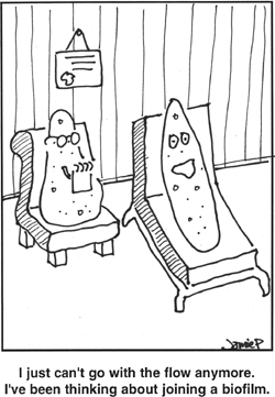
Who would guess that studying ancient artwork offers an excellent opportunity to learn about bacterial metabolism? Bacteria degrade art and historic treasures in the same way they decompose organic matter. Bacterial enzymes lipase and protease break down fats and peptides, respectively, in pigments and a variety of carbohydrate and fiber-degrading enzymes attack the canvas and wood. These are the same enzymes animals use for digesting food. Different, more specialized bacteria get energy from chemical reactions involving inorganic salts. All of these microbial actions contribute to the slow decomposition of the world’s greatest works of art, principally because of bacteria acting in concert within a community (see Figure 4.1).
Figure 4.1. Biofilm bugs. (Courtesy of Center for Biofilm Engineering, Montana State University)
The decomposition of artwork is one small piece of bacterial cycling of nutrients on Earth. Bacteria circulate the Earth’s elements through the atmosphere, water, soil, and plant and animal life in processes called nutrient or biogeochemical cycles. These cycles take place in the seas, forests, and mountains. Nutrient cycling also occurs when bacteria decompose rubber, plastic water bottles, paint, and countless manmade items that were once believed to be indestructible. Bacteria corrode metal, stone, marble, and concrete, and they degrade paint, paper, canvas, leather, pigments, and wood. Chemical reactions driven by bacteria weaken modern infrastructure such as bridges, roadways, and oil tankers. In exactly the same way, bacteria have been steadily digesting the components found in art, whether these are made of metal, fiber, hide, or pigments.
Copper is one of the oldest metals used in civilization. In the Bronze Age (3000-1300 BCE) craftsmen took advantage of the metal’s malleability to incorporate it into the alloys brass and bronze for tools, weapons, domestic items (bowls, plates, goblets, and so on), and jewelry. Microbiologists have learned just in the past several years that sulfate-reducing bacteria have been corroding bronzes such as Etruscan relics dating to the ninth century BCE. When a bacterium is said to “reduce” an element, it means a bacterial enzyme adds electrons to the element. In metal corrosion, sulfate-reducing bacteria convert sulfate, a sulfur atom with oxygens attached, to the element sulfur. When a biofilm forms on relics containing iron, the anaerobic bacteria at the base of the film convert sulfur to pyrite, an iron atom attached to two sulfurs. Figure 4.2 illustrates the substantial biofilm growth that metal structures can support.
Figure 4.2. Biofilm corrosion. This pipe has been almost completely occluded by biofilm, which has dried and hardened. Few technologies exist for removing biofilm from living or nonliving surfaces.
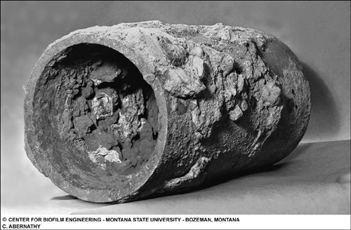
(Courtesy of Center for Biofilm Engineering, Montana State University)
Sulfate-reducing Desulfovibrio and the iron-oxidizing Leptothrix work in concert to corrode iron; they are sometimes nicknamed “iron-eating” bacteria. Leptothrix takes electrons away from iron atoms at the metal’s surface, and Desulfovibrio, hovering a few μm nearby, accepts the excess electrons. Even though iron corrodes when exposed to the air, the biofilm actually creates tiny anaerobic nooks called microenvironments in which these reactions take place. Sergei Winogradsky discovered the general steps in iron-sulfur metabolism between 1885 and 1889.
Bacterial deterioration of metal takes place every day 12,850 feet at the bottom of the Atlantic. The H.M.S. Titanic has withstood the incredible hydrostatic pressures exerted on it for a century. The oxygenless deep waters have also allowed the ship to resist rusting. Yet the Titanic supports a ghostly collection of rusticles. These appendages from several inches to a few feet long and numbering in the thousands hang from almost every part of the ship. Some are as fragile as tissue; others hold their shape when research vessels pull them to the surface. The rusticles demonstrate that the main cause for the Titanic’s inexorable return to the Earth is bacteria.
The rusticles contain a mixture of bacteria able to thrive in the cold and deep where the Titanic’s wreck settled on April 15, 1912. The bacteria remove 0.3 gram of iron from every square centimeter of the ship daily. The loss of iron causes about 300 kilograms of steel to detach from the wreck each day. Anaerobic “iron-eating” bacteria have been taking apart the Titanic one iron atom at a time and may cause the hull to cave in on itself within 100 years, perhaps as little as 40 years from now. The ship’s organic material, mostly wood paneling and fixtures, serve as the main nutrient source for the bacteria, but as the metals corrode more organic matter may be exposed. As a result, the deterioration of the Titanic will accelerate.
Stone and concrete undergo similar aboveground weathering. The corrosion of ancient Greek and Roman stone statues illustrate bacteria’s role in the slowest of all biogeochemical cycles, the rock cycle or sediment cycle. Unlike nitrogen, which can migrate from soil to the atmosphere and return to plants within a day, the rock cycle takes eons to complete. Bacteria begin by weakening the stone through corrosion. Small pieces break off from the mass and make their way downhill with erosion. Erosion carries the matter to bodies of water where it sinks and becomes part of sediment. Sediments, especially under the ocean, compact under intense pressure. As tectonic plates shift, this sediment becomes part of metamorphic rock in the Earth’s mantle and slowly pushes upward to the planet’s surface. Some metamorphic rock sinks into the Earth’s interior where the planet’s molten core heats the sediment and turns it into magma. Magma rushes to the surface all at once in volcanoes. New rock that has either migrated up to the Earth’s surface gradually or exploded toward the surface from a volcano becomes available for bacteria to again begin degrading.
Molecular methods have now been applied to studies of how bacteria affect not only rock, but also prehistoric paintings in moist caves. The 20,000-year-old cave paintings in Altamira, Spain, and Lascaux, France, may be succumbing to the combined activities of bacteria that degrade the dyes as well as the underlying stone. Many of the offending bacteria have not yet been identified, but microbiologists have noticed that Actinobacteria often dominate the microbial populations in the Spanish and French caves. Actinobacteria build tentacles called filaments that grow into the pores of rock surfaces, allowing the bacterial damage to occur in the rock’s subsurface. Molecular analyses of the cave paintings have also uncovered aerobes (Pseudomonas) and anaerobes (Thiovulum), sulfate- (Desulfovibrio) and iron-users (Shewanella), bacteria that use a wide variety of nutrients (Clostridium), and species with narrow nutrient requirements (Thiobacillus). Lascaux’s 600 paintings made of a mixture of mineral pigments and animal fat offers a banquet for the bacteria of the caves.
Art galleries have almost as difficult a time protecting treasures from bacterial decay, despite humidity and temperature-controlled environments. Fungi and the funguslike bacterium Actinomyces extend filaments into paintings’ surfaces to cause physical destruction. Other microbes chemically decompose the pigments. Polymerase chain reaction (PCR) technology, invented in the 1980s, has enabled microbiologists to multiply bits of bacterial DNA recovered from painted frescoes on castles in Austria, Germany, and France to study the types and proportions of the bacteria. The analyses have so far revealed the presence of Clostridium, Frankia, and Halomonas on the ceiling painting in Castle Herberstein in Styria, Austria. Each genus contributes its own mode of destruction on the painting:
• Clostridium—A spore-forming anaerobe that grows well on a wide variety of chemicals
• Frankia—A spore-forming genus that builds long, branching filaments that penetrate surfaces
• Halomonas—A versatile halophile that lives with or without oxygen and can degrade alcohols, acids, and organic solvents
The constant streams of tourists who visit the world’s great works of art accelerate decay. Human bodies and breath change the temperature and humidity in galleries and even in caves containing ancient wall paintings. The Lascaux caves had been in good condition when discovered in 1940. The rapid decay of the cave paintings that took place after people started visiting then led to the closure of Lascaux in 1965 to prevent further deterioration.
On stone exposed to the weather, biofilms and cyanobacteria each contribute in their own way to the deterioration of statues, buildings, and headstones. In some instances, bacterial growth on historical structures presents no more than a cosmetic problem due to the discoloration of stone by bacterial pigments. In other cases, acids produced by bacteria in biofilms degrade the stone’s calcium carbonate as they degrade tooth enamel in dental caries. The actions of fungi, biofilm bacteria, and free bacteria with their different types of metabolism hasten the decomposition of historical structures and have already decimated most concrete structures, not only from ancient periods but since the 20th century.
Lichens form greenish black stains on old stone structures. Lichen is a living entity made from a cooperative relationship between a fungus and a bacterium or an alga. Among bacteria, cyanobacteria are by far the most common to form associations with fungi. The photosynthesis performed by cyanobacteria helps keep the lichen alive but also may aid the growth of other bacteria by providing organic nutrients. The limestone surfaces of the Mayan ruins at Chichen-Itza support bacteria and other microbes. The bacterial populations become denser and more diverse on the sun-bathed stones and less dense and varied on surfaces inside temples and corridors.
Microbiologists have taken a unique tack for preventing the further deterioration of art by using other bacteria to combat the effects of art-degrading species. The first task involves cleaning the objects to remove the heavy detritus that has accumulated over centuries. Specific bacteria remove crusts of sulfates and nitrates, animal-based glues, and the remains of molds and insects. Injecting nutrients into the pores of art’s materials might allow bacteria to grow and form crystals that would block future infiltration of the porous surfaces. Similar bacteria may be applied to clean the microscopic crevices of a piece. Researcher Giancarlo Ranalli, working in Pesche, Italy, has become an expert on using bacteria to clean art. He has applied bacteria-filled poultices to clean the marble of Michelangelo’s Pieta Rondanini, and in 2007 his team reported a comparison between bacteria and a cleaning mixture of ammonium carbonate, detergent, and an abrasive applied to marble surfaces in Milan Cathedral. Desulfovibrio vulgaris cleaned the marble without removing the material’s patina while the cleaning mixture removed less dirt and left behind a precipitate. Ranalli’s team has since put Pseudomonas stutzeri to work digesting glue from protective shrouds that had covered frescoes in the Pisa Cemetery for more than 20 years. In this case, P. stutzeri’s unique protein-degrading enzymes released the fabric without destroying the fresco underneath.
Conservators of Europe’s galleries have been hesitant to let a microbiologist spread bacteria all over their valuable works of art. Although bacteria like Ranalli’s have worked on stone, the same method does not have a long track record on gallery art. Cleaning a 300-year-old painting differs from using bacteria to dissolve crud from inside restaurant grease traps, septic tanks, and wastewater holding tanks in navy ships, all current uses for Bacillus, Pseudomonas, and other bacteria. Art-cleaning bacteria will require fine-tuning to ensure they digest unwanted dirt but leave the art’s components in good condition.
Biotechnology might help in the new field of bacteria-based art refurbishing. Bacteria can be engineered to excrete antibiotics targeted to art-loving bacteria. Genetically modified organisms (GMOs) might also someday act on specific components in art buildup, and then stop due to a shut-off gene that activates when the target compounds are gone. In the meanwhile, bacteria gnaw on the Roman Coliseum and perhaps the Mona Lisa.