22

Multifunctional scaffolds for bone tissue engineering and in situ drug delivery

V. Mouriño and J.P. Cattalini,    University of Buenos Aires, Argentina

W. Li and A.R. Boccaccini,    University of Erlangen-Nuremberg, Germany

S. Lucangioli,    University of Buenos Aires, Argentina

Abstract:

This chapter provides an overview about the development of bone tissue engineering scaffolds with the ability to provide the controlled delivery of therapeutic drugs. Typical drugs considered include gentamicin and other antibiotics generally used to combat osteomyelitis as well as anti-inflammatory drugs and bisphosphonates. Special attention has been given to the technology used for controlling the release of the loaded drugs. A detailed summary of drugs included in bone tissue scaffolds is presented and the many approaches developed to combine organic and inorganic biomaterials in composites for drug-delivery systems are discussed. The remaining challenges in the field are summarized, suggesting also future research directions.

Key words

multifunctional scaffolds; drug delivery; bone tissue engineering; therapeutic drugs

22.1 Introduction

Although bone tissue has the unique ability to heal, repair and remodel, there is a growing need to tackle several debilitating and deadly conditions, due to trauma or disease, which require the replacement of the affected bone (Buckwalter et al. 1996a, 1996b). In addition the increasing aging population is leading to more requirements for the effective treatment of bone defects and the need to accelerate the healing of large bone fractures. Tissue engineering (TE) offers an alternative to donor graft tissue (autografts, allografts, or xenografts) for the treatment of bone diseases (Goessler et al. 2007; Lee and Shin 2007; Bran et al. 2008; Kanczler and Oreffo 2008; Guarino et al. 2012; Nguyen et al., 2012). In this sense, bone TE can potentially improve the lives of those patients who are suffering from bone diseases associated with tissue loss by providing a controlled environment that promotes and directs cell attachment, proliferation and differentiation, and for supporting new tissue growth (Mouriño et al. 2013). A common approach to bone tissue engineering involves the application of biodegradable and biocompatible scaffolds to create in the first instance sufficient space for new tissue formation, e.g. a three-dimensional (3D) engineered porous biomaterials which must: (a) promote the infiltration and proliferation of host cells for tissue regeneration (Duarte et al. 2007), (b) exhibit high porosity, high pore interconnectivity and uniform pore distribution to enable vascularization (Lee and Shin 2007; Cipitria et al. 2012; Guarino et al. 2012; Nguyen et al. 2012) and (c) have the ability to be resorbed at a rate similar to the rate of new tissue formation. Therefore, the selection of the most appropriate biomaterials for the preparation of scaffolds should be done considering the mechanical and physicochemical requirements as well as their degree of biocompatibility and absence of adverse immune responses (Hutmacher 2000; Mouriño et al. 2013). The desired characteristics of traditional scaffolds for bone TE (BTE) and their fabrication technologies have been described in several review articles (for example in Garg et al., 2012; Blackwood et al., 2012; Hutmacher, 2000; Guarino et al., 2007; Rezwan et al., 2006). In addition, scaffolds have to be suitable for sterilization without losing their properties.

A convenient alternative being increasingly investigated to improve the scaffold biological functionality is to load therapeutic drugs in scaffolds to support the treatment of bone disorders and/or to combat possible infections. Such drugs incorporated into tissue scaffolds must be released with an adequate therapeutic concentration level and for a desired time frame (Gomes and Reis 2004; Duarte et al. 2007; Baroli 2009; Mouriño and Boccaccini 2010; Hafeman et al. 2010; Wende and Guelcher 2011). The increasing research activities associated with this approach, which promotes the development of matrices with a dual function: scaffolds for the growth of new tissue and carriers for controlled drug delivery in situ, is leading to the novel research field called TE therapeutic (Baroli 2009; Mouriño and Boccaccini 2010). In this chapter, which follows from our previous review paper (Mouriño and Boccaccini 2010), special attention has been paid to the latest developments related to control the release rate of relevant drugs from bone TE scaffolds based on different organic and inorganic biomaterials. However, the development of bone TE scaffolds with the specific capability to deliver growth factors or other bioactive molecules, being a very important subtopic in bone TE strategies (Wende and Guelcher 2011; Vo et al. 2012; Ekenseair et al. 2013), will not be covered in this chapter. The document is organized as follows: Section 22.2 is dedicated to summarizing the biomaterials utilized and scaffold designs proposed as drug delivery vehicles. Section 22.3 details several approaches adopted to develop bone TE scaffolds with drug delivery capability considering antimicrobial agents, anti-inflammatory and antiresorptive drugs. Finally, the remaining challenges in the field are summarized in Section 22.4, where also directions for future research efforts are highlighted.

22.2 Scaffolds as drug carriers

Scaffolds for bone TE are made from a variety of biodegradable polymers, bioactive and resorbable inorganic materials including calcium phosphates, mesoporous silica and bioactive glasses, and their composites, as discussed in several chapters of this book. There are increasing investigations focusing on loading engineered scaffolds with therapeutic drugs, generating a dual function for the matrices: (i) scaffolds in the ‘classical’ TE approach, e.g. for the delivery of cells and to support the growth of new tissue (Langer and Vacanti 1993; Freed et al. 1994; Bonassar and Vacanti 1998; Shea et al. 1999; Hutmacher 2000; Hoffman 2002; Guarino et al. 2012) and (ii) carriers for controlled in situ drug delivery (Drury and Mooney 2003; Sokolsky-Papkov et al. 2007; Kretlow and Mikos 2008; Lyons et al. 2008; Makarov et al. 2010; Kankilic et al. 2011; Lee et al. 2011; Wende and Guelcher 2011; Hum and Boccaccini 2012). Therapeutic drugs used in the treatment of bone diseases administered locally have several advantages over systemic administration such as the reduction of adverse effects and the risk of overdose, while enhancing the bioavailability of the drug with the appropriate therapeutic concentration effectively reaching the target site (Baroli 2010; Mouriño and Boccaccini 2010; Mouriño et al. 2013).

In order to design and develop multifunctional scaffolds, several variables must be taken into account. Generally, multifunctional scaffolds must enable the delivery of therapeutic drugs to the nearby tissues, and must provide adequate control of the rate of release of the loaded drugs in order to sustain the expected concentration level in their target sites for the required period of time. The selection of processing methods to develop highly porous scaffolds with suitable mechanical and structural properties is one of the challenges in the field of bone TE (Hutmacher 2000; Rezwan et al. 2006; Guarino et al. 2012; Liu et al. 2013). The effect of the drug incorporation on the physicochemical and mechanical properties of scaffolds as well as the controlled release of drugs from the matrices must be taken into account when multifunctional scaffolds are designed. From a pharmaceutical standpoint, the type of interaction between drug and scaffold, the mechanism of degradation of the scaffold and the selection of the fabrication process will be determined by the type of drug, its stability and the required release kinetics. Further, the level and duration of the therapeutic drug may need to be modulated to avoid excessive drug activity at the target site and it may also be useful a drug delivery with time varying concentrations.

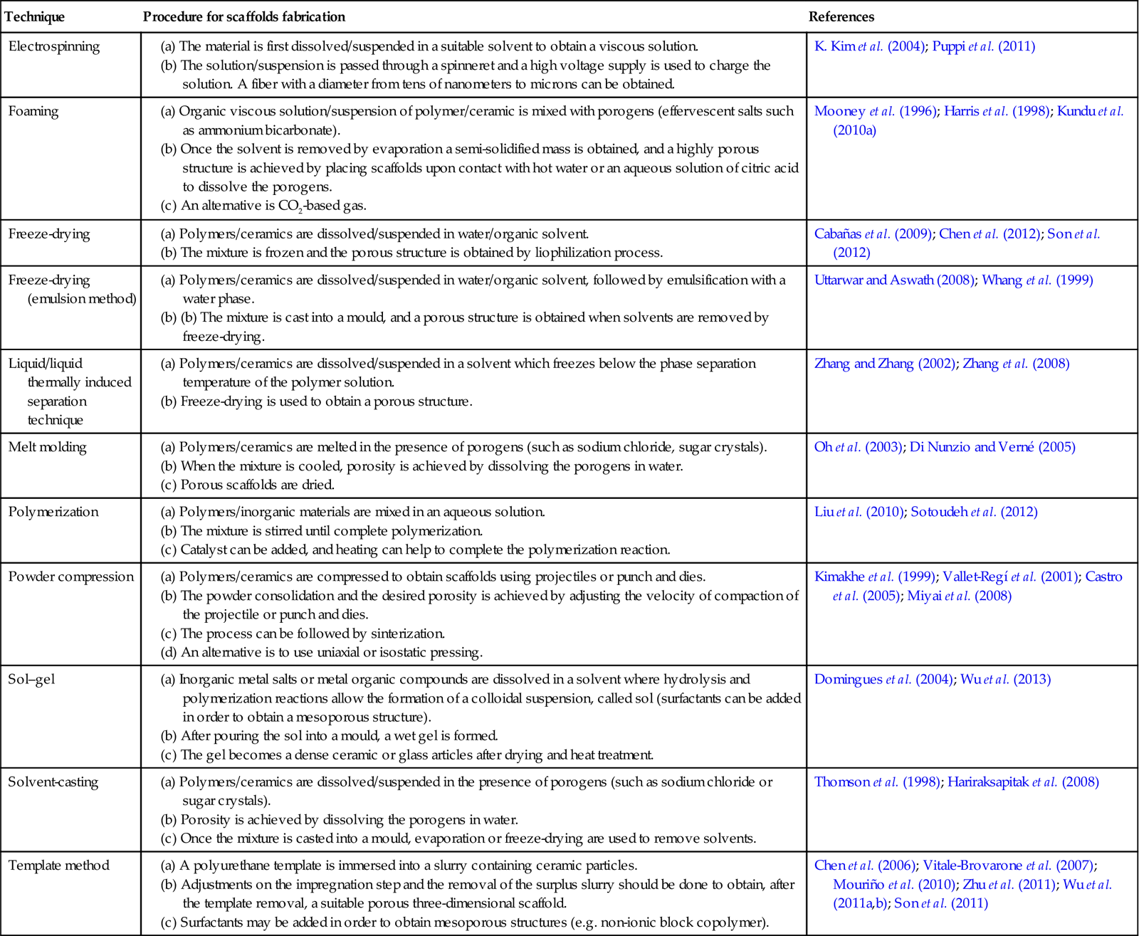
Processing methods for TE scaffolds usually involve processes that are incompatible with the incorporation and stability of organic drugs (Drury and Mooney 2003). Fabrication conditions such as high temperatures, use of certain organic solvents, application of pressure and free radicals may lead to drug decomposition and should be avoided (Mandal and Kundu 2008). The techniques more frequently used to fabricate bioactive bone scaffolds with potential drug release capability are listed in Table 22.1 and a summary of the evolution of experimental research carried out on the development of 3D scaffolds for bone tissue engineering with controlled release capability is schematized in Fig. 22.1. These scaffolds can be developed with the potential to provide not only the physicochemical environment and the structural integrity required for bone regeneration (the main scaffold function), but also with the added function of local regulator to control the dose and kinetics of drug release effectively acting as the drug carrier (Berger et al. 1997; Mouriño and Boccaccini 2010).

Table 22.1

Different techniques to elaborate porous three-dimensional scaffolds with drug-delivery capability for bone tissue engineering therapeutics

Technique Procedure for scaffolds fabrication References
Electrospinning

(a) The material is first dissolved/suspended in a suitable solvent to obtain a viscous solution.

(b) The solution/suspension is passed through a spinneret and a high voltage supply is used to charge the solution. A fiber with a diameter from tens of nanometers to microns can be obtained.

K. Kim et al. (2004); Puppi et al. (2011)
Foaming

(a) Organic viscous solution/suspension of polymer/ceramic is mixed with porogens (effervescent salts such as ammonium bicarbonate).

(b) Once the solvent is removed by evaporation a semi-solidified mass is obtained, and a highly porous structure is achieved by placing scaffolds upon contact with hot water or an aqueous solution of citric acid to dissolve the porogens.

(c) An alternative is CO2-based gas.

Mooney et al. (1996); Harris et al. (1998); Kundu et al. (2010a)
Freeze-drying

(a) Polymers/ceramics are dissolved/suspended in water/organic solvent.

(b) The mixture is frozen and the porous structure is obtained by liophilization process.

Cabañas et al. (2009); Chen et al. (2012); Son et al. (2012)
Freeze-drying (emulsion method)

(a) Polymers/ceramics are dissolved/suspended in water/organic solvent, followed by emulsification with a water phase.

(b) (b) The mixture is cast into a mould, and a porous structure is obtained when solvents are removed by freeze-drying.

Uttarwar and Aswath (2008); Whang et al. (1999)
Liquid/liquid thermally induced separation technique

(a) Polymers/ceramics are dissolved/suspended in a solvent which freezes below the phase separation temperature of the polymer solution.

(b) Freeze-drying is used to obtain a porous structure.

Zhang and Zhang (2002); Zhang et al. (2008)
Melt molding

(a) Polymers/ceramics are melted in the presence of porogens (such as sodium chloride, sugar crystals).

(b) When the mixture is cooled, porosity is achieved by dissolving the porogens in water.

(c) Porous scaffolds are dried.

Oh et al. (2003); Di Nunzio and Verné (2005)
Polymerization

(a) Polymers/inorganic materials are mixed in an aqueous solution.

(b) The mixture is stirred until complete polymerization.

(c) Catalyst can be added, and heating can help to complete the polymerization reaction.

Liu et al. (2010); Sotoudeh et al. (2012)
Powder compression

(a) Polymers/ceramics are compressed to obtain scaffolds using projectiles or punch and dies.

(b) The powder consolidation and the desired porosity is achieved by adjusting the velocity of compaction of the projectile or punch and dies.

(c) The process can be followed by sinterization.

(d) An alternative is to use uniaxial or isostatic pressing.

Kimakhe et al. (1999); Vallet-Regí et al. (2001); Castro et al. (2005); Miyai et al. (2008)
Sol–gel

(a) Inorganic metal salts or metal organic compounds are dissolved in a solvent where hydrolysis and polymerization reactions allow the formation of a colloidal suspension, called sol (surfactants can be added in order to obtain a mesoporous structure).

(b) After pouring the sol into a mould, a wet gel is formed.

(c) The gel becomes a dense ceramic or glass articles after drying and heat treatment.

Domingues et al. (2004); Wu et al. (2013)
Solvent-casting

(a) Polymers/ceramics are dissolved/suspended in the presence of porogens (such as sodium chloride or sugar crystals).

(b) Porosity is achieved by dissolving the porogens in water.

(c) Once the mixture is casted into a mould, evaporation or freeze-drying are used to remove solvents.

Thomson et al. (1998); Hariraksapitak et al. (2008)
Template method

(a) A polyurethane template is immersed into a slurry containing ceramic particles.

(b) Adjustments on the impregnation step and the removal of the surplus slurry should be done to obtain, after the template removal, a suitable porous three-dimensional scaffold.

(c) Surfactants may be added in order to obtain mesoporous structures (e.g. non-ionic block copolymer).

Chen et al. (2006); Vitale-Brovarone et al. (2007); Mouriño et al. (2010); Zhu et al. (2011); Wu et al. (2011a,b); Son et al. (2011)

Image

image
22.1 Summary of the evolution of experimental research carried out on the development of 3D scaffolds for bone tissue engineering with controlled release capability.

22.3 Controlled release of therapeutic drugs for bone tissue engineering

Different strategies have been proposed to enable the release in a controlled manner of relevant therapeutic drugs for the treatment of diseases associated with bone repair process in 3D scaffolds used in bone TE. A detailed summary of the most common drugs loaded in such scaffolds and both in vitro and in vivo studies carried out is presented in Table 22.2. Typical drugs considered include gentamicin and other antibiotics generally used to combat osteomyelitis such as tetracycline, polymyxin B, gatifloxacine and ciprofloxacine; as well as silver, anti-inflammatory drugs and bisphosphonates. From Table 22.2 it appears that porous matrices based mainly on well-characterized biocompatible polymeric scaffolds or, in some cases, composites comprising polymeric matrices and added inorganic particles, represent convenient systems to incorporate therapeutic drug delivery in bone TE approaches. A schematic diagram summarizing the different strategies proposed is shown in Plate XIV (between pages 354 and 355). Although several novel techniques have been developed to introduce therapeutic drugs within scaffolds, in most cases the strategy followed has been the direct incorporation of the drug into the scaffold by immersion of the scaffold in a drug containing buffer aqueous solution. Nevertheless, thermo-labile drugs can also be loaded within 3D scaffolds in a one step process using room temperature compaction of powder mixtures, this being a solvent-free process which avoids the use of toxic solvents (Kimakhe et al. 1999; Vallet-Regí et al. 2001; Castro et al. 2005). Alternatively drug carriers in the form of biodegradable polymer microspheres can be loaded into the 3D scaffold structure (Francis et al. 2010). Such a structure is shown in Fig 22.2 where drug loaded P3HB microspheres have been deposited on the surface of bioactive glass scaffolds following an approach similar to that developed by Francis et al. (2010). Generally, it is observed that the release kinetics of drugs loaded in multifunctional scaffolds is not necessarily directly linked to the degradation kinetic of the biodegradable scaffold. Even though most of the 3D scaffolds reviewed in this chapter have been shown to have rates of degradation much lower than the required rate of drug release, it is important to highlight that drug release from most of the developed scaffolds is mainly driven by the process of diffusion through them. Moreover, few studies are focused on delivering the drugs within specific therapeutic levels over a predetermined period of time and taken into account the particular in vivo microenvironment including the effect of vascularization.

Table 22.2

Examples of drug delivery from three-dimensional scaffolds for bone tissue engineering

Therapeutic effect Therapeutic drug Matrix composition Matrix shape Process technique Type of Experimental trial References
Antibiotic/antibacterial Amoxicillin Nano zeolite/PEG/poly acrylic acid/polyacrylamid Composite pieces Nano zeolite was added to a mixture containing polymers and drug + stirring until complete polymerization in vitro Sotoudeh et al. (2012)
 Ceftazidime EC microspheres/HA/PU Porous matrix A mixture of HA/PEG/catalyst was stirred under dry nitrogen atmosphere + a chain extender was added and the temperature was maintained at 65 °C + an aqueous suspension of drug-loaded microspheres was added with a catalyst + the mixture was poured into a mold at 80 °C to complete the polymerization + washing + drying in vitro Liu et al. (2010)
 Ceftriaxone/sulbactam HA/β-TCP/chitosan Porous matrix Foaming method in vitro Kundu et al. (2010b)
 Ciproflozacin HA/β-TCP/PLA Porous matrix Compression in vitro/in vivo Castro et al. (2005)
 Colistin PLGA microspheres/PMMA/CMC Porous construct The mixture of loaded-PLGA microspheres + PMMA + CMC was cast in molds and allowed to harden in vitro Shi et al. (2010)
 Copper Bioactive glass/alginate Porous matrix Polyurethane sponge technique + immersion in alginate solution + crosslinking in vitro Erol et al. (2012)
  Mesoporous bioactive glass Porous matrix Sol–gel in vitro Wu et al. (2013)
 Gallium Alginate/bioactive glass Porous matrix Bioactive glass scaffold made by foam replica technique + scaffold coating with gallium crosslinked alginate in vitro Mouriño et al. (2010)
  Alginate/bioactive glass nanoparticles Films The mixture containing alginate and bioactive glass nanoparticles was casted into a mould and left to dry + crosslinking with gallium in vitro Mouriño et al. (2011)
 Gatifloxacin β-TCP/PCL Porous matrix Compaction + sintering + immersion in drug-loaded PCL slurry in vitro/in vivo Miyai et al. (2008)
 Gentamicin β-TCP/CP/chitosan Porous matrix Thermally induced phase-separation technique + immersion in drug-containing PBS solution in vitro Zhang and Zhang (2002)
  Bioactive glass Bioactive glass pieces Uniaxial and isostatic compression at room temperature in vivo Vallet-Regí et al. (2001)
Antibiotic/antibacterial  Bioactive glass Mesoporous bioactive glass/bioactive glass Polyurethane sponge technique + immersion in drug-containing PBS solution in vitro Zhu and Kaskel (2009)
  Bioactive glass/P(3HB) microspheres Porous matrix Polyurethane sponge technique + emulsion solvent evaporation method to obtain gentamicin-loaded microspheres + immersion in microsphere slurry in vitro Francis et al. (2010)
  HMS-HA/PLGA microspheres Porous matrix Tensioactive template to obtain HMS-HA + double-emulsion evaporation technique to obtain GS-loaded PLGA microspheres + sinterization at 70 °C in vitro Shi et al. (2009)
  Zirconium/bioactive glass Mesoporous bioactive glass Polyurethane sponge technique + evaporation-induced self-assembly process + immersion in drug-containing solution in vitro Zhu et al. (2011)
 Polymyxin B Calcium phosphate Ceramic pieces Compaction in vitro Kimakhe et al. (1999)
 Silver Bioactive glass Bioactive glass pieces Melting and sintering at high temperature + ion-exchange process to introduce the drug in vitro Di Nunzio and Verné (2005)
 Tetracycline Bioactive glass/β-cyclodextrin Bioactive glass pieces Sol–gel in vivo Domingues et al. (2004)
  Chitosan/HA Porous matrix Freeze-drying in vitro Teng et al. (2009)
  HA/PCL Porous matrix Polyurethane sponge technique + immersion in drug/HA/PCL slurry in vitro H. Kim et al. (2004c)
 Vancomycin β-TCP/agarose Porous matrix Freeze-drying and heat desiccation at 37 °C. in vitro Cabañas et al. 2009)
  (Gelatin/β-TCP Porous matrix A mixture of gelatin/β-TCP/drug/genipin was stirred + gelification + crosslinking + freeze-drying in vivo Zhou et al. (2012)
  HA/PCL Porous matrix Polyurethane sponge technique + immersion in drug/HA/PCL slurry in vitro Kim et al. (2005)
  PDLLA/BCP/alginate Porous matrix Particle: leaching/thermally induced phase separation method + immersion in alginate/vancomycin solution + crosslinking in vitro Zhang et al. (2008)
Antibiotic/antitumoral Doxorubicin PCL/chitosan/nanoclay/β-TCP Porous matrix PCL scaffolds were made by fused deposition modeling. The clay/DOX carrier was added to a chitosan/β-TCP mixture. Finally, PCL scaffolds were submerged into the mentioned mixture and freeze dried. in vitro Chen et al. (2012)
Antiinflammatory Ibuprofen Mesoporous bioactive glass Porous matrix Sol–gel + immersion in ibuprofen-hexane solution in vitro Wu et al. (2013)
  HA Porous matrix Cellulose sponge technique + immersion in drug solution in vitro Palazzo et al. 2005)
Inductive effect in osteogenic culture Dexamethasone Starch/PLA Porous matrix Supercritical phase-inversion technique in vitro Duarte et al. (2009a)
  Chitosan Porous matrix Freeze-drying + drug impregnation by supercritical fluid technology in vitro Duarte et al. 2009b)
  Boron + bioactive glass Mesoporous bioactive glass Co-templates of nonionic block polymer + polyurethane sponge technique + immersion in drug-containing PBS solution in vitro Wu et al. (2011a)
  SiO2 Mesoporous structure Co-templates of nonionic block polymer + polyurethane sponge technique + immersion in drug-containing PBS solution in vitro Wu et al. (2011b)
  Drug-loaded PLGA nanoparticles/HA Porous matrix Polymeric template coating technique in vitro/in vivo Son et al. (2011)
  Drug-loaded PLGA nanoparticles/HA/TCP Granules Freeze-drying in vitro Son et al. (2012)
Inhibition of the osteoclastic resorption Alendronate PLGA/HA Microspheric scaffold in vitro Wang et al. (2010)
  Poly(L-lactide-co-epsilon caprolactone)/bioactive glass Microspheric scaffold Emulsion-solvent + evaporation method in vitro Mondal et al. (2012)
  Silica (SBA-15) Mesoporous silica matrix Triblock copolymers technique + functionalization + immersion in drug-containing buffer aqueous solution in vitro Nieto et al. (2008)
Inhibition of the osteoclastic resorption Clodronate PCL/HA Porous matrix Drug-loaded HA nanoparticles were added to a PCL solution + fibers obtained by electrospinning in vitro Puppi et al. (2011)
 Pamidronate PDLLA Pellets Solvent casting method + compression in vivo Yu et al. (2010)
 Zoledronate CPA Pellets Suspension of CDA in drug containing water solution in vitro Faucheux et al. (2009)
Ibuprofen Bioactive glass/MCM-41 Porous matrix MCM-41 spheres + melting and sitering at high temperature to obtain bioactive glass scaffold + drug impregnation in vitro Mortera et al. (2010)

Image

Image

b-TCP, b-tricalcium phosphate; CPA, calcium phosphate-deficient apatite; CMC, carboxymethylcellulose; CP, calcium phosphate invert glasses; ES, ethyl cellulose; GS, gentamicin; HA, hydroxyapatite; HMS, mesoporous silica; PCL, poly-1-caprolactone; PBS, phosphate-buffered saline; PCL, poly(ε-caprolactone); PDLLA, poly(D,L-lactic acid); PEG, poly(ethylene glycol); PLA, poly(L-lactic acid); PLGA, poly(lactide-co-glycolide); PMMA, polymethylmethacrylate; PU, polyurethane.

image
22.2 Scanning electron microscopy (SEM) image showing the structure of the strut of a Bioglass®-based scaffold coated with drug loaded P(3HB) microspheres fabricated by the technique developed by Francis et al. (2010).

In addition, in most cases the effect of the formation of apatite surface layer and precipitation of hydroxyapatite, which is common in bioactive bone tissue scaffolds, on drug release kinetics has not been contemplated during in vitro release studies.

22.4 Conclusions and future trends

Research carried out, particularly in recent years, on bioactive bone TE scaffolds with additional drug-delivery capability has indicated the great potential of such multifunctional scaffolds for application in bone re-generation. Especially encouraging are the results obtained with combinations of materials, as well as improved 3D bone tissue scaffold designs based on novel processing techniques with the added value of drug-delivery capability. However, and despite the significant progress achieved, there is still the need to fully understand the correlation between in vitro and in vivo performance of the designed scaffolds, and probably it will take time to achieve in vivo results of relevance which can provide a rational for optimization of drug-delivery function of multifunctional scaffolds.

Further efforts should be made in developing strategies to establish the concentration and distribution of a therapeutic drug within a scaffold which is required for successful outcomes. At present, most of the investigated approaches present reduced ability to adjust drug dosages and most of the systems developed would not be easily scalable for commercial applications in terms of cost effectible manufacture process. In this sense, multifunctional scaffolds should be easy to produce, sterilize and handle. Finally, another important consideration, often overlooked, is the requirements needed to obtain approval from regulatory authorities.

It is clear that, in order to optimize these systems, various disciplines such as chemistry, biology, pharmacy, medicine and biomaterials science must come together in interdisciplinary approaches. The interdisciplinary character of TE – allowing the confluence of different scientific fields, backgrounds and knowledge – is mandatory for the great deal of further work required towards the development of more effective multifunctional bone TE.

22.5 References and further reading

1. Ahmad Z, Zhang H, Farook U, Edirisinghe M, Stride E, Colombo P. Generation of multi-layered structures for biomedical applications using a novel trineedle coaxial device and electrohydrodynamic flow. J R Soc Interface. 2008;5:1255–1261. doi 10.1098/rsif.2008.0247.

2. Ambrose C, Gogola G, Clyburn T, Raymond A, Peng A, Mikos A. Antibiotic microspheres: preliminary testing for potential treatment of osteomyelitis. Clin Orthop Relat Res. 2003;415:279–285. doi 10.1097/01.blo.0000093920.26658.ae.

3. Aoyagi S, Onishi H, Machida Y. Novel chitosan wound dressing loaded with minocycline for the treatment of severe burn wounds. Int J Pharm. 2007;330:138–145. doi 10.1016/j.ijpharm.2006.09.016.

4. Baroli B. From natural bone graft to tissue engineering therapeutics: brainstorming on pharmaceutical formulative requirements and challenges. J Pharm Sci. 2009;98:1317–1375. doi 10.1002/jps.21528.

5. Berens A, Huvard G, Korsmeyer R, Kunig F. Application of compressed carbon dioxide in the incorporation of additives into polymers. J Appl Polym Sci. 1992;46:231–242. doi 10.1002/app.1992.070460204.

6. Berger R, Jacobs J, Quigley L, Rosenberg A, Galante J. Primary cementless acetabular reconstruction in patients younger than 50 years old 7- to 11-year results. Clin Orthop Relat Res. 1997;344:216–226.

7. Blackwood K, Bock N, Dargaville Y, Woodruff MA. Scaffolds for growth factor delivery as applied to bone tissue engineering. Int J Polym Sci 2012. doi 10.1155/2012/174942.

8. Bonassar J, Vacanti C. Tissue engineering: the first decade and beyond. J Cell Biochem Suppl. 1998;30:297–303.

9. Bostman O, Pihlajamaki H. Clinical biocompatibility of biodegradable orthopaedic implants for internal fixation: a review. Biomaterials. 2000;21:2615–2621. doi 10.1016/S0142-9612(00)00129-0.

10. Bran G, Stern-Straeter J, Hormann K, Riedel F, Goessler U. Apoptosis in bone for tissue engineering. Arch Med Res. 2008;39:467–482. doi 10.1016/j.arcmed.2008.02.007.

11. Buckwalter J, Glimcher M, Cooper R, Recker R. Bone biology I Structure, blood supply, cells, matrix, and mineralization Instr. Course Lect. 1996a;45:371–386.

12. Buckwalter J, Glimcher M, Cooper R, Recker R. Bone biology II Formation, form, modeling, remodeling, and regulation of cell function Instr. Course Lect. 1996b;45:387–399.

13. Cabañas M, Peña J, Román J, Vallet-Regí M. Tailoring vancomycin release from x-TCP/agarose scaffolds. Eur J Pharm Sci. 2009;37:249–256. doi 10.1016/j.ejps.2009.02.011.

14. Castro C, Sánchez E, Delgado A, et al. Ciprofloxacin implants for bone infection In vitro–in vivo characterization. J Control Release. 2003;93:341–354. doi 10.1016/j.jconrel.2003.09.004.

15. Castro C, Évora C, Baro M, Soriano I, Sánchez E. Two-month ciprofloxacin implants for multibacterial bone infections. Eur J Pharm Sci. 2005;60:401–406.

16. Chen M, Le D, Hein S, et al. Fabrication and characterization of a rapid prototyped tissue engineering scaffold with embedded multicomponent matrix for controlled drug release. Int J Nanomedicine. 2012;7:4285–4297. doi 10.2147/IJN.S33083.

17. Chen Q, Thompson I, Boccaccini A. 45S5 Bioglass-derived glass ceramic scaffolds for bone tissue engineering. Biomaterials. 2006;27:2414–2425. doi 10.1016/j.biomaterials.2005.11.025.

18. Chung L, Schmidt R, Hamlyn P, Sagar B, Andrews A, Turner T. Biocompatibility of potential wound management products: fungal mycelia as a source of chitin/chitosan and their effect on the proliferation of human F1000 fibroblasts in culture. J Biomed Mater Res. 1994;24:463–469.

19. Cipitria A, Lange C, Schell H, et al. Porous scaffold architecture guides tissue formation. J Bone Miner Res. 2012;27:1275–1288. doi 10.1002/jbmr.1589.

20. Day R, Boccaccini A, Shurey S, et al. Assessment of polyglycolic acid mesh and bioactive glass for soft tissue engineering scaffolds. Biomaterials. 2004;25:5857–5866. doi 10.1016/j.biomaterials.2004.01.043.

21. Di Nunzio S, Verné E. In: Process for the production of silver-containing prosthetic devices. 2005; Italian Patent no. PCT/EP2005/056391.

22. Di Nunzio S, Vitale-Brovarone C, Spriano S, et al. Silver containing bioactive glasses prepared by molten salt ion exchange. J Eur Ceram Soc. 2004;24:2935–2942.

23. Domingues Z, Cortés M, Gomes T, et al. Bioactive glass as a drug delivery system of tetracycline and tetracycline associated with β-cyclodextrin. Biomaterials. 2004;25:327–333. doi 10.1016/S0142-9612(03)00524-6.

24. Dong J, Kojima H, Uemura T, Kikuchi M, Tateishi T, Tanaka J. In vivo evaluation of a novel porous hydroxyapatite to sustain osteogenesis of transplanted bone marrow-derived osteoblastic cells. J Biomed Mater Res. 2001;57:208–216. doi 10.1002/1097-4636(200111)57:2,208:AID-JBM1160.3.0.CO;2-N.

25. Drury J, Mooney D. Hydrogels for tissue engineering: scaffold design variables and applications. Biomaterials. 2003;24:4337–4351.

26. Duarte A, Simplicio A, Vega-González A, et al. Supercritical fluid impregnation of a biocompatible polymer for ophthalmic drug delivery. J Supercritical Fluids. 2007;42:373–377. doi 10.1016/j.supflu.2007.01.007.

27. Duarte A, Mano J, Reis R. Dexamethasone-loaded scaffolds prepared by supercritical-assisted phase inversion. Acta Biomater. 2009b;5:2054–2062. doi 10.1016/j.actbio.2009.01.047.

28. Duarte A, Mano J, Reis R. Preparation of chitosan scaffolds loaded with dexamethasone for tissue engineering applications using supercritical fluid technology. Eur Polym J. 2009a;45:141–148. doi 10.1016/j.eurpolymj.2008.10.004.

29. Ekenseair A, Kasper F, Mikos A. Perspectives on the interface of drug delivery and tissue engineering. Adv Drug Deliv Rev. 2013;65:89–92. doi 10.1016/j.addr.2012.08.017.

30. Erol M, Ozyuguran A, Ozarpat O, Kucukbayrak S. 3D composite scaffolds using strontium containing bioactive glasses. J Eur Ceram Soc. 2012;32:2747–2755. doi 10.1016/j.jeurceramsoc.2012.01.015.

31. Faucheux C, Verron E, Soueidan A, et al. Controlled release of bisphosphonate from a calcium phosphate biomaterial inhibits osteoclastic resorption in vitro. J Biomed Mater Res Part A. 2009;86:46–56.

32. Francis L, Meng D, Knowles J, Roy I, Boccaccini AR. Multifunctional P(3HB) microsphere/45S5 Bioglass-based composite scaffolds for bone tissue engineering. Acta Biomater. 2010;6:2773–2786. doi 10.1016/j.actbio.2009.12.054.

33. Freed L, Vunjak-Novakovic G, Biron R, et al. Biodegradable polymer scaffolds for tissue engineering. Nat Biotechnol. 1994;12:689–693.

34. Freyman M, Yannas YV, Gibson L. Cellular materials as porous scaffolds for tissue engineering. J Prog Mater Sci. 2001;46:273–282. doi 10.1016/S0079-6425(00)00018-9.

35. Fujibayashi S, Neo M, Kim HM, Kokubo T, Nakamura T. A comparative in vivo bone ingrowth and in vitro apatite formation on Na2O–CaO–SiO2 glasses. Biomaterials. 2003;24:1349–1356. doi 10.1016/S0142-9612(02)00511-2.

36. Garg T, Singh O, Arora S, et al. Scaffold: a novel carrier for cell and drug delivery. Crit Rev Ther Drug Carrier Syst. 2012;29:1–63.

37. Garvin K, Miyano J, Robinson D, Giger D, Novak J, Radio S. Polylactide/polyglycolide antibiotic implants in the treatment of osteomyelitis. A canine model J Bone Joint Surg Am. 1994;76:1500–1506.

38. Goessler U, Stern-Straeter J, Riedel K, Bran G, Hormann K, Riedel F. Tissue engineering in head and neck reconstructive surgery: what type of tissue do we need? Eur Arch Otorhinolaryngol. 2007;264:1343–1356. doi 10.1007/s00405-007-0369-y.

39. Gomes M, Reis R. Biodegradable polymers and composites in biomedical applications from catgut to tissue engineering Part II Systems for temporary replacement and advanced tissue regeneration. Int Mater Rev. 2004;49:274–285. doi 10.1179/095066004225021927.

40. Guarino V, Causa F, Ambrosio L. Bioactive scaffolds for bone and ligament tissue. Exp Rev Med Devices. 2007;4:405–418. doi 10.1586/17434440.4.3. 405.

41. Guarino V, Gloria A, Raucci M, De Santis R, Ambrosio L. Bio-inspired composite and cell instructive platforms for bone regeneration. Int Mater Rev. 2012;57:256–275.

42. Gupta A, Seifalian A, Edirisinghe M, Winslet M. Novel electrohydrodynamic printing of nanocomposite biopolymer scaffolds. J Bioactive Compatible Polym. 2007;22:265–280. doi 10.1177/0883911507078268.

43. Habraken W, Wolke J, Jansen J. Ceramic composites as matrices and scaffolds for drug delivery in tissue engineering. Adv Drug Deliv Rev. 2007;59:234–248. doi 10.1016/j.addr.2007.03.011.

44. Hafeman A, Zienkiewicz K, Carney E, et al. Local delivery of tobramycin from injectable biodegradable polyurethane scaffolds. J Biomater Sci Polym Ed. 2010;21:95–112. doi 10.1163/156856209X410256.

45. Han D, Gouma P. Electrospun bioscaffolds that mimic the topology of extracellular matrix. Nanomed Nanotechnol Biol Med. 2006;2:37–41. doi 10.1016/j.nano.2006.01.002.

46. Hariraksapitak P, Suwantong O, Pavasant P, Supaphol P. Effectual drug-releasing porous scaffolds from 1,6-diisocyanatohexane-extended poly (1,4-butylene succinate) for bone tissue regeneration. Polymer. 2008;49:2678–2685. doi 10.1016/j.polymer.2008.04.006.

47. Harris L, Kim B, Mooney D. Open pore biodegradable matrices formed with gas foaming. J Biomed Mater Res. 1998;42:396–402. doi 10.1002/(SICI)1097-4636(19981205)42:3,396:AID-JBM7.3.0.CO;2-E.

48. Heller J. Controlled drug release from poly(ortho esters)-a surface eroding polymer. J Control Release. 1985;2:167–177. doi 10.1016/0168-3659(85)90042-2.

49. Hench L. Bioceramics J Am Ceram Soc. 1998;81:1705–1728.

50. Hendricks K, Lane D, Burd T, et al. Elution characteristics of tobramycin from polycaprolactone in a rabbit model. Clin Orthop Relat Res. 2001;392:418–426. doi 10.1097/00003086-200111000-00055.

51. Heyde M, Partridge K, Howdle S, Oreffo R, Garnett M, Shakesheff K. Development of a controlled non-viral DNA release system from PDLLA scaffolds fabricated using a supercritical CO2 technique. Biotechnol Bioeng. 2007;98:679–693. doi 10.1002/bit.21446.

52. Hoffman A. Hydrogels for biomedical applications. Adv Drug Deliv Rev. 2002;43:3–12.

53. Hollister S. Porous scaffold design for tissue engineering. Nat Mater. 2005;4:518–524. doi 10.1038/nmat1421.

54. Hum J, Boccaccini A. Bioactive glasses as carriers for bioactive molecules and therapeutic drugs: a review. J Mater Sci Mater Med. 2012;23:2317–2333.

55. Hutmacher D. Scaffolds in tissue engineering bone and cartilage. Biomaterials. 2000;21:2529–2543.

56. Hutmacher D. Scaffold design and fabrication technologies for engineering tissues: state of the art and future perspectives. J Biomater Sci Polym Ed. 2001;12:107–124. doi 10.1163/156856201744489.

57. Hutmacher D, Cool S. Concepts of scaffold-based tissue engineering: the rationale to use solid free-form fabrication techniques. J Cell Mol Med. 2007;11:654–669. doi 10.1111/j.1582-4934.2007.00078.x.

58. Hutmacher D, Schantz J, Lam C, Tan K, Lim T. State of the art and future directions of scaffold-based bone engineering from a biomaterials perspective. J Tissue Eng Regen Med. 2007;1:245–260. doi 10.1002/term.24.

59. Izquierdo-Barba I, Monserrat C, Vallet-Regí M. Nanostructured mesoporous silicas for bone tissue regeneration. J Nanomater. 2008;3:1–14.

60. Jacob E, Setterstrom J, Bach D, Heath III J, McNiesh L, Cierny III G. Evaluation of biodegradable ampicillin anhydrate microcapsules for local treatment of experimental staphylococcal osteomyelitis. Clin Orthop Relat Res. 1991;267:237–244.

61. Kanczler J, Oreffo R. Osteogenesis and angiogenesis: the potential for engineering bone. Eur Cell Mater. 2008;15:100–114.

62. Kanellakopoulou K, Giamarellos-Bourboulis E. Carrier systems for the local delivery of antibiotics in bone infections. Drugs. 2000;59:1223–1232. doi 10.2165/00003495-200059060-00003.

63. Kankilic B, Bayramli E, Kilic E, Dağdeviren S, Korkusuz F. Vancomycin containing PLLA/β-TCP controls MRSA in vitro. Clin Orthop Relat Res. 2011;469:3222–3228. doi 10.1007/s11999-011-2082-9.

64. Karageorgiou V, Kaplan D. Porosity of 3D biomaterial scaffolds and osteogenesis. Biomaterials. 2006;26:5474–5491. doi 10.1016/j.biomaterials.2005.02.002.

65. Kazarian S. Polymer processing with supercritical fluids. Polym Sci Ser C. 2000;42:78–101.

66. Kikic I, Sist P. Applications of supercritical fluids to pharmaceuticals: controlled drug delivery systems, supercritical fluids: fundamentals and applications in. In: Proc. Second NATO ASI on Supercritical Fluids, NATO Science Series. Dordrecht, The Netherlands: Kluwer Academic Publishers; 2000:291–306.

67. Kim H, Miyaji F, Kokubo T, Ohtsuki C, Nakamura T. Bioactivity of Na2O–CaO–SiO2 glasses. J Am Ceram Soc. 1995;78:2405–2411. doi 10.1111/j.1151-2916.1995.tb08677.x.

68. Kim H, Knowles J, Kim H. Development of hydroxyapatite bone scaffold for controlled drug release via poly(1-caprolactone) and hydroxyapatite hybrid coatings. J Biomed Mater Res Part B: Appl Biomater. 2004a;70:240–249.

69. Kim H, Knowles J, Kim E. Hydroxyapatite and gelatine composite foams processed via novel freeze-drying and crosslinking for use as temporary hard tissue scaffolds. J Biomed Mater Res A. 2004b;72:136–145.

70. Kim H, Knowles J, Kim H. Hydroxyapatite/poly(ε-caprolactone) composite coatings on hydroxyapatite porous bone scaffold for drug delivery. Biomaterials. 2004c;25:1279–1287.

71. Kim H, Knowles J, Kim H. Hydroxyapatite porous scaffold engineered with biological polymer hybrid coating for antibiotic Vancomycin release. J Mater Sci Mater Med. 2005;16:189–195. doi 10.1007/s10856-005-6679-y.

72. Kim K, Luu Y, Chang C, et al. Incorporation and controlled release of a hydrophilic antibiotic using poly(lactide-co-glycolide)-based electrospun nanofibrous scaffolds. J Control Release. 2004d;98:47–56. doi 10.1016/j.jconrel.2004.04.009.

73. Kimakhe S, Bohic S, Larosse C, et al. Biological activities of sustained polymixin B release from calcium phosphate biomaterial prepared by dynamic compaction: an in vitro study. J Biomed Mater Res. 1999;47:18–27. doi 10.1002/(SICI)1097-4636(199910)47:1,18:AID-JBM3.3.0.CO;2-T.

74. Kretlow J, Mikos A. From material to tissue: biomaterial development, scaffold fabrication, and tissue engineering. AIChE J. 2008;54:3048–3067.

75. Kundu B, Soundrapandian C, Nandi SK, et al. Development of new localized drug delivery system based on ceftriaxone-sulbactam composite drug impregnated porous hydroxyapatite: a systematic approach for in vitro and in vivo animal trial. Pharm Res. 2010a;27:1659–1676.

76. Kundu B, Lemos A, Soundrapandian C, et al. Development of porous HAp and β-TCP scaffolds by starch consolidation with foaming method and drug-chitosan bilayered scaffold based drug delivery system. Journal of Materials Science: Materials in Medicine. 2010b;21:2955–2969.

77. Langer R, Vacanti J. Tissue engineering. Science. 1993;260:920–926.

78. Lee G, Park J, Shin U, Kim H. Direct deposited porous scaffolds of calcium phosphate cement with alginate for drug delivery and bone tissue engineering. Acta Biomater. 2011;7:3178–3186.

79. Lee J, Park J, Kwang H, Sang H, Kim J, Hwal S. An infection-preventing bilayered collagen membrane containing antibiotic-loaded hyaluronan microparticles: physical and biological properties. Artif Organs. 2002;26:636–646. doi 10.1046/j.1525-1594.2002.06847.x.

80. Lee S, Shin H. Matrices and scaffolds for delivery of bioactive molecules in bone and cartilage tissue engineering. Adv Drug Deliv Rev. 2007;59:339–359. doi 10.1016/j.addr.2007.03.016.

81. Li L, Deng J, Stephens D. Polyanhydride implant for antibiotic delivery: from the bench to the clinic. Adv Drug Deliv Rev. 2002a;54:963–986.

82. Li W, Laurencin C, Caterson E, Tuan R, Ko F. Electrospun nanofibrous structure: a novel scaffold for tissue engineering. J Biomed Mater Res. 2002b;60:613–621. doi 10.1002/jbm.10167.

83. Liu H, Zhang L, Shi P, Zou Q, Zuo Y, Li Y. Hydroxyapatite/polyurethane scaffold incorporated with drug-loaded ethyl cellulose microspheres for bone regeneration. J Biomed Mater Res B Appl Biomater. 2010;95:36–46. doi 10.1002/jbm.b.31680.

84. Liu Y, Lim J, Teoh S. Review: Development of clinically relevant scaffolds for vascularised bone tissue engineering. Biotechnol Adv. 2013;31:688–705 (doi:pii:S0734-9750(12)00184-X.10.1016/j.biotechadv.2012.10.003).

85. Livingston T, Ducheyne P, Garino J. In vivo evaluation of a bioactive scaffold for bone tissue engineering. J Biomed Mater Res. 2002;62:1–13. doi 10.1002/jbm.10157.

86. Lu L, Garcia CA, Mikos AG. In vitro degradation of thin poly(DL-lactic-co-glycolic acid) films. J Biomed Mater Res. 1999;46:236–244. doi 10.1002/(SICI)1097-4636(199908)46:2,236:AID-JBM13.3.0.CO;2-F.

87. Lyons F, Partap S, O’Brien F. Part 1: scaffolds and surfaces. Technol Health Care. 2008;16:305–317.

88. Ma Z, Kotaki M, Inai R, Ramakrishna S. Potential of nanofiber matrix as tissue-engineering scaffolds. Tissue Eng. 2005;11:101–109. doi 10.1089/ten.2005.11.101.

89. Makarov C, Gotman I, Radin S, Ducheyne P, Gutmanas E. Vancomycin release from bioresorbable calcium phosphate-polymer composites with high ceramic volume fractions. J Mater Sci Mater Med. 2010;45:6320–6324.

90. Malafaya P, Silva G, Baran E, Reis R. Drug delivery therapies II: strategies for delivering bone regenerating factors. Curr Opin Solid State Mater Sci. 2002;6:297–312. doi 10.1016/S1359-0286(02)00077-3.

91. Mandal B, Kundu S. Non-bioengineered high strength three-dimensional gland fibroin scaffolds from tropical non-mulberry silkworm for potential tissue engineering applications. Macromol Biosci. 2008;8:807–818.

92. Matthews J, Wnek G, Simpson D, Bowlin G. Electrospinning of collagen nanofibers. Biomacromolecules. 2002;3:232–238. doi 10.1021/bm015533u.

93. Melville A, Rodriguez-Lorenzo L, Forsythe J. Effects of calcination temperature on the drug delivery behaviour of Ibuprofen from hydroxyapatite powders. J Mater Sci Mater Med. 2008;19:1187–1195. doi 10.1007/s10856-007-3185-4.

94. Mi F, Wu Y, Shyu S, et al. Control of wound infections using a bilayer chitosan wound dressing with sustainable antibiotic delivery. J Biomed Mater Res. 2002;59:438–449. doi 10.1002/jbm.1260.

95. Misra S, Valappil S, Roy I, Boccaccini A. Polyhydroxyalkanoate (PHA)/inorganic phase composites for tissue engineering applications. Biomacromolecules. 2006;7:2249–2258. doi 10.1021/bm060317c.

96. Misra S, Ansari T, Valappil S, et al. Poly(3-hydroxybutyrate) multifunctional composite scaffolds for tissue engineering applications. Biomaterials. 2010;31:2806–2815. doi 10.1016/j.biomaterials.2009.12.045.

97. Miyai T, Ito A, Tamazawa G, et al. Antibiotic-loaded poly-e-caprolactone and porous β-tricalcium phosphate composite for treating osteomyelitis. Biomaterials. 2008;29:350–358. doi 10.1016/j.biomaterials.2007.09.040.

98. Mondal T, Sunny M, Khastgir D, Varma H, Ramesh P. Poly (L-lactide-co-epsilon caprolactone) microspheres laden with bioactive glass-ceramic and alendronate sodium as bone regenerative scaffolds. Mater Sci Eng C Mater Biol Appl. 2012;32:697–706. doi 10.1016/j.msec.2012.01.011.

99. Mooney D, Baldwin D, Suh N, Vacanti J, Langer R. Novel approach to fabricate porous sponges of poly(d, l-lactic-co-glycolic acid) without the use of organic solvents. Biomaterials. 1996;17:1417–1422. doi 10.1016/0142-9612(96)87284-X.

100. Mortera R, Onida B, Fiorilli S, et al. Synthesis and characterization of MCM-41 spheres inside bioactive glass-ceramic scaffold. Chem Eng J. 2007;137:54–61. doi 10.1016/j.cej.2007.07.094.

101. Mortera R, Baino F, Croce G, et al. Monodisperse mesoporous silica spheres inside a bioactive macroporous glass–ceramic scaffold. Adv Eng Mater. 2010;12:B256–B259. doi 10.1002/adem.200980075.

102. Mouriño V, Boccaccini A. Bone tissue engineering therapeutics: controlled drug delivery in three-dimensional scaffolds. J R Soc Interface. 2010;7:209–227. doi 10.1098/rsif.2009.0379.

103. Mouriño V, Newby P, Boccaccini A. Preparation and characterization of gallium releasing 3-D alginate coated 4555 Bioglass® based scaffolds for bone tissue engineering. Adv Eng Mater. 2010;12:8283–8291. doi 10.1002/adem.200980078.

104. Mouriño V, Newby P, Pishbin P, Cattalini J, Lucangioli S, Boccaccini A. Physicochemical, biological and drug-release properties of gallium crosslinked alginate/nanoparticulate bioactive glass composite films. Soft Matter. 2011;7:6705–6712. doi 10.1039/C1SM05331K.

105. Mouriño V, Cattalini JP, Roether JA, Dubey P, Roy I, Boccaccini AR. Composite polymer-bioceramic scaffolds with drug delivery capability for bone tissue engineering. Expert Opin Drug Deliv. 2013;10(10):1353–1365. doi 10.1517/17425247.2013.808183.

106. Muzzarelli R, Tarsi R, Filippini O, Giovanetti E, Biagini G, Varaldo P. Antimicrobial properties of Ncarboxybutyl chitosan. Antimicrob Agents Chemother. 1990;34:2019–2023.

107. Nair L, Laurencin C. Biodegradable polymers as biomaterials. Prog Polym Sci. 2007;32:762–798. doi 10.1016/j.progpolymsci.2007.05.017.

108. Nelson C, Hickmon S, Skinner R. Treatment of experimental osteomyelitis by surgical debridement and the implantation of bioerodable, polyanhy-dridegentamicin beads. J Orthop Res. 1997;15:249–255. doi 10.1002/jor.1100150214.

109. Nguyen L, Annabi N, Nikkhah M, et al. Vascularized bone tissue engineering: approaches for potential improvement. Tissue Eng Part B Rev. 2012;18:363–382.

110. Nicoll S, Radin S, Santos E, Tuan R, Ducheyne P. In vitro release kinetics of biologically active transforming growth factor-x1 from a novel porous glass carrier. Biomaterials. 1997;18:853–859. doi 10.1016/S0142-9612(97)00008-2.

111. Nie L, Nicolau D, Nightingale C, Browner B, Quintilliani R. In vitro elution of ofloxacin from a bioabsorbable polymer. Acta Orthop Scand. 1995;66:365–368.

112. Nieto A, Balas F, Colilla M, Manzano M, Vallet-Regí M. Functionalization degree of SBA-15 as key factor to modulate sodium alendronate dosage. Microporous Mesoporous Mater. 2008;116:4–13. doi 10.1016/j.micromeso.2008.03.025.

113. Oh S, Kang S, Kim E, Cho S, Lee J. Fabrication and characterization of hydrophilic poly(lactic-co-glycolic acid)/poly(vinyl alcohol) blend cell scaffolds by melt-molding particulate-leaching method. Biomaterials. 2003;24:4011–4021. doi 10.1016/s0142-9612(03)00284-9.

114. Overbeck J, Winckler S, Meffert R, Tormala P, Spiege H, Brug E. Penetration of ciprofloxacin into bone: a new bioabsorbable implant. J Invest Surg. 1995;8:155–162. doi 10.3109/08941939509023138.

115. Palazzo B, Sidoti M, Roveri N, et al. Controlled drug delivery from porous hydroxyapatite grafts: an experimental and theoretical approach. Mater Sci Eng. 2005;25:207–213. doi 10.1016/j.msec.2005.01.011.

116. Pang L, Hu Y, Yan Y, et al. Surface modification of PLGA/s-TCP scaffold for bone tissue engineering: hybridization with collagen and apatite. Surf Coat Technol. 2007;201:9549–9557. doi 10.1016/j.surfcoat.2007.04.035.

117. Park S, Kim J, Suh H. Evaluation of antibiotic-loaded collagen-hyaluronic acid matrix as a skin substitute. Biomaterials. 2004;25:3689–3698. doi 10.1016/j.biomaterials.2003.10.072.

118. Prabu P, Dharmaraj N, Aryal S, Lee B, Ramesh V, Kim H. Preparation and drug release activity of scaffolds containing collagen and poly(caprolactone). J Biomed Mater Res A. 2006;79:153–158.

119. Price J, Tencer A, Arm D, Bohach G. Controlled release of antibiotics from coated orthopaedic implants. J Biomed Mater Res. 1996;30:281–286. doi 10.1002/(SICI)1097-4636(199603)30:3,281:AID-JBM2.3.0.CO;2-M.

120. Puppi D, Piras A, Chiellini F, et al. Optimized electro- and wet-spinning techniques for the production of polymeric fibrous scaffolds loaded with bisphosphonate and hydroxyapatite. J Tissue Eng Regen Med. 2011;5:253–263. doi 10.1002/term.310.

121. Rai R, Keshavarz T, Roether J, Boccaccini A, Roy I. Medium chain length polyhydroxyalkanoates, promising new biomedical materials for the future. Mater Sci Eng R Rep. 2011;72:29–47. doi 10.1016/j.mser.2010.11.002.

122. Ramay H, Zhang M. Preparation of porous hydroxyapatite scaffolds by combination of the gel-casting and polymer sponge methods. Biomaterials. 2003;24:3293–3302. doi 10.1016/S0142-9612(03)00171-6.

123. Rezwan K, Chen Q, Blaker J, Boccaccini A. Biodegradable and bioactive porous polymer/inorganic composite scaffolds for bone tissue engineering. Biomaterials. 2006;27:3413–3431. doi 10.1016/j.biomaterials.2006.01.039.

124. Roether A, Boccaccini A, Hench L, Maquet V, Gautier S, Jérome R. Development and in vitro characterisation of novel bioresorbable and bioactive composite materials based on polylactide foams and Bioglass for tissue engineering applications. Biomaterials. 2002;23:3871–3878. doi 10.1016/S0142-9612(02)00131-X.

125. Roman J, Madruga E. Polymers with pharmacological activity: synthesis and free radical polymerization of p-methacryloyloxyacetanilide. Polymer. 1989;30:949–954. doi 10.1016/0032-3861(89)90198-5.

126. Rossi S, Azghani A, Omri A. Antimicrobial efficacy of a new antibiotic-loaded poly(hydroxybutyricco-hydroxyvaleric acid) controlled release system. J Antimicrob Chemother. 2004;54:1013–1018. doi 10.1093/jac/dkh477.

127. Rossi S, Marciello M, Sandri G, et al. Wound dressings based on chitosans and hyaluronic acid for the release of chlorhexidine diacetate in skin ulcer therapy. Pharm Dev Technol. 2007;12:415–422. doi 10.1080/10837450701366903.

128. Ruszczak Z, Friess W. Collagen as a carrier for on-site delivery of antibacterial drugs. Adv Drug Deliv Rev. 2003;55:1679–1698. doi 10.1016/j.addr.2003.08.007.

129. Rutledge B, Huyette D, Day D, Anglen J. Treatment of osteomyelitis with local antibiotics delivered via bioabsorbable polymer. Clin Orthop Relat Res. 2003;411:280–287. doi 10.1097/01.blo.0000065836.93465.ed.

130. Santos E, Radin S, Ducheyne P. Sol–gel derived carrier for the controlled release of proteins. Biomaterials. 1999;20:1695–1700. doi 10.1016/S0142-9612(99)00066-6.

131. Shanmugasundaram N, Sundaraseelan J, Uma S, Selvaraj D, Babu M. Design and delivery of silver sulfadiazine from alginate microspheres-impregnated collagen scaffold. J Biomed Mater Res B Appl Biomater. 2006;77:378–388.

132. Shea L, Smiley E, Bonadio J, Mooney D. DNA delivery from polymer matrices for tissue engineering. Nat Biotechnol. 1999;17:551–554.

133. Shi M, Kretlow J, Nguyen A, et al. Antibiotic-releasing porous polymethylmethacrylate constructs for osseous space maintenance and infection control. Biomaterials. 2010;31:4146–4156. doi 10.1016/j.biomaterials.2010.01.112.

134. Shi X, Wang Y, Rena L, Zhao N, Gong Y, Wang D. Novel mesoporous silica-based antibiotic releasing scaffold for bone repair. Acta Biomater. 2009;5:1697–1707. doi 10.1016/j.actbio.2009.01.010.

135. Sokolsky-Papkov M, Agashi K, Olaye A, Shakesheff K, Domb A. Polymer carriers for drug delivery in tissue engineering. Adv Drug Deliv Rev. 2007;59:187–206.

136. Somayaji B, Jariwala U, Jayachandran P, Vidyalakshmi K, Dudhani R. Evaluation of antimicrobial efficacy and release pattern of tetracycline and metronidazole using a local delivery system. J Periodontol. 1998;69:409–413.

137. Son J, Appleford M, Ong J, et al. Porous hydroxyapatite scaffold with three-dimensional localized drug delivery system using biodegradable microspheres. J Control Release. 2011;153:133–140. doi 10.1016/j.jconrel.2011.03.010.

138. Son J, Lee K, Kim S, Kwon T, Kim K. Porous calcium phosphate granules containing drug-loaded polymeric nanoparticles for bone regeneration. Mater Lett. 2012;76:243–246. doi 10.1016/j.matlet.2012.02.114.

139. Sotoudeh S, Barati A, Davarnejad R, Aliabadi Farahani M. Antibiotic release process from hydrogel nano zeolite composites. Middle-East J Sci Res. 2012;12:392–396. doi 10.5829/idosi.mejsr.2012.12.3.1062.

140. Sripriya R, Kumar M, Sehgal P. Improved collagen bilayer dressing for the controlled release of drugs. J Biomed Mater Res B Appl Biomater. 2004;70:389–396.

141. Streubel J, Siepmann J, Peppas N, Bodmeier R. Bimodal drug release achieved with multi-layer matrix tablets: transport mechanisms and device design. J Control Release. 2000;69:455–468. doi 10.1016/S0168-3659(00)00334-5.

142. Teng S, Lee E, Wang P, Jun S, Han C, Kim H. Functionally gradient chitosan/hydroxyapatite composite scaffolds for controlled drug release. J Biomed Mater Res B Appl Biomater. 2009;90:275–282. doi 10.1002/jbm.b.31283.

143. Thomson R, Yaszemski M, Powers J, Mikos A. Hydroxyapatite fiber reinforced poly(a-hydroxylester) foams for bone regeneration. Biomaterials. 1998;18:1935–1943.

144. Turesin F, Gursel I, Hasirci V. Biodegradable polyhydroxyalkanoate implants for osteomyelitis therapy: in vitro antibiotic release. J Biomater Sci Polym Ed. 2001;12:195–207. doi 10.1163/156856201750180924.

145. Uttarwar M, Aswath P. Fabrication of porous, drug-releasing, biodegradable, polymer scaffolds for sustained drug release. J Biomed Mater Res B Appl Biomater. 2008;87:121–131. doi 10.1002/jbm.b.31077.

146. Vallet-Regí M, Ragel C, Arcos D, Clavel-Sainz M, Meseguer-Olmo L. In: Métodos para la obtención de implantes bioactivos útiles como sistemas de liberación controlada. 2001; Spanish Patent no. P22001–01386.2181593.

147. Vallet-Regí M, Ruiz-González L, Izquierdo-Barba I, González-Calbet J. Revisiting silica based ordered mesoporous materials: medical applications. J Mater Chem. 2006;16:26–31. doi 10.1039/b509744d.

148. Vallet-Regí M, Balas F, Colilla M, Manzano M. Bone-regenerative bioceramic implants with drug and protein controlled delivery capability. Prog Solid State Chem. 2008;36:163–191.

149. Vitale-Brovarone C, Di Nunzio S, Bretcanu O, Verné E. Macroporous glass-ceramic materials with bioactive properties. J Mater Sci. Mater Med. 2004;15:209–217. doi 10.1023/B:JMSM.0000015480.49061.e1.

150. Vitale-Brovarone C, Verné E, Appendino P. Macroporous bioactive glass-ceramic scaffolds for tissue engineering. J Mater Sci. Mater Med. 2006;17:1069–1078. doi 10.1007/s10856-006-0533-8.

151. Vitale-Brovarone C, Verné E, Robiglio L, et al. Development of glass–ceramic scaffolds for bone tissue engineering: characterisation, proliferation of human osteoblasts and nodule formation. Acta Biomater. 2007;2:199–208.

152. Vitale-Brovarone C, Miola M, Balagna C, Verné E. 3D-glass–ceramic scaffolds with antibacterial properties for bone grafting. Chem Eng J. 2008;137:129–136. doi 10.1016/j.cej.2007.07.083.

153. Vo T, Kasper F, Mikos A. Strategies for controlled delivery of growth factors and cells for bone regeneration. Adv Drug Deliv Rev. 2012;64:1292–1309. doi 10.1016/j.addr.2012.01.016 Epub 2012 Feb 4.

154. Wang H, Li Y, Zuo Y, Li J, Ma S, Cheng L. Biocompatibility and osteogenesis of biomimetic nanohydroxyapatite/polyamide composite scaffolds for bone tissue engineering. Biomaterials. 2007;28:3338–3348. doi 10.1016/j.biomaterials.2007.04.014.

155. Wang Y, Shi X, Ren L, Yao Y, Wang D. In vitro osteogenesis of synovium mesenchymal cells induced by controlled release of alendronate and dexamethasone from a sintered microspherical scaffold. J Biomater Sci Polym Ed. 2010;21:1227–1238. doi 10.1163/092050609x12481751806259.

156. Wende J, Guelcher S. Dual delivery of an antibiotic and a growth factor addresses both the microbiological and biological challenges of contaminated bone fractures. Expert Opin Drug Deliv. 2011;8:1555–1569.

157. Whang K, Healy K, Elenz D, et al. Engineering bone regeneration with bioabsorbable scaffolds with novel microarchitecture. Tissue Eng. 1999;5:35–51.

158. Wu C, Miron R, Sculean A, et al. Proliferation, differentiation and gene expression of osteoblasts in boron-containing associated with dexamethasone deliver from mesoporous bioactive glass scaffolds. Biomaterials. 2011a;32:7068–7078. doi 10.1016/j.biomaterials.2011.06.009.

159. Wu C, Fan W, Chang J, Xiao Y. Mussel-inspired porous SiO2 scaffolds with improved mineralization and cytocompatibility for drug delivery and bone tissue engineering. J Mater Chem. 2011b;21:18300–18307. doi 10.1039/c1jm12770e.

160. Wu Ch, Zhou Y, Xu M, et al. Copper-containing mesoporous bioactive glass scaffolds with multifunctional properties of angiogenesis capacity, osteostimulation and antibacterial activity. Biomaterials. 2013;34:422–433.

161. Wu P, Grainger D. Drug/device combinations for local drug therapies and infection prophylaxis. Biomaterials. 2006;27:2450–2467. doi 10.1016/j.biomaterials.2005.11.031.

162. Xu Q, Czernuszka J. Controlled release of amoxicillin from hydroxyapatite coated poly(lactic-co-glycolic acid) microspheres. J Control Release. 2008;127:146–153.

163. Xynos I, Edgar A, Buttery L, Hench L. Gene-expression profiling of human osteoblasts following treatment with the ionic products of Bioglass 45S5 dissolution. J Biomed Mater Res. 2001;55:151–157. doi 10.1002/1097-4636(200105)55:2,151:AID-JBM1001.3.0.CO;2-D.

164. Yagmurlu M, Korkusuz F, Gursel I, Korkusuz P, Ors U, Hasirci V. Sulbactam-cefoperazone polyhydroxybutyrate-co-hydroxyvalerate (PHBV) local antibiotic delivery system: in vivo effectiveness and biocompatibility in the treatment of implant-related experimental osteomyelitis. J Biomed Mater Res. 1999;46:494–503. doi 10.1002/(SICI)1097-4636(19990915)46:4,494:AID-JBM7.3.0.CO;2-E.

165. Yu N, Schindeler A, Peacock L, et al. In vivo local co-delivery of recombinant human bone morphogenetic protein-7 and pamidronate via poly-d, l-lactic acid. Eur Cell Mater. 2010;20:431–442.

166. Yuan H, Kurashina K, Beuijn J, Li Y, De Groot K, Zhang X. A preliminary study on osteoinduction of two kinds of calcium phosphate ceramics. Biomaterials. 1999;20:1799–1806. doi 10.1016/S0142-9612(99)00075-7.

167. Yunos D, Bretcanu O, Boccaccini A. Polymer-bioceramic composites for tissue engineering scaffolds. J Mater Sci. 2008;43:4433–4442.

168. Zhang C, Wang J, Feng H, Lu B, Song Z, Zhang X. Replacement of segmental bone defects using porous bioceramic cylinders: a biomechanical and X-ray diffraction study. J Biomed Mater Res. 2001;54:407–411. doi 10.1002/1097-4636(20010305)54:3,407:AID-JBM140.3.0.CO;2-C.

169. Zhang L, Yang D, Chen H, et al. An ionically crosslinked hydrogel containing vancomycin coating on a porous scaffold for drug delivery and cell culture. Int J Pharm. 2008;353:74–87. doi 10.1016/j.ijpharm.2007.11.023.

170. Zhang Y, Zhang M. Calcium phosphate/chitosan composite scaffolds for controlled in vitro antibiotic drug release. J Biomed Mater Res. 2002;62:378–386. doi 10.1002/jbm.10312.

171. Zhou J, Fang T, Wang Y, Dong J. The controlled release of vancomycin in gelatin/beta-TCP composite scaffolds. J Biomed Mater Res A. 2012;100:2295–2301. doi 10.1002/jbm.a.34170.

172. Zhu Y, Kaskel S. Comparison of the in vitro bioactivity and drug release property of mesoporous bioactive glasses (MBGs) and bioactive glasses (BGs) scaffolds. Microporous Mesoporous Mater. 2009;118:176–182. doi 10.1016/j.micromeso.2008.08.046.

173. Zhu Y, Zhang Y, Wu Ch, Fang Y, Yang J, Wang S. The effect of zirconium incorporation on the physiochemical and biological properties of mesoporous bioactive glasses scaffolds. Microporous and Mesoporous Materials. 2011;143:311–319.

174. Zilberman M, Elsner J. Antibiotic-eluting medical devices for various applications. J Control Release. 2008;130:202–215. doi 10.1016/j.jconrel.2008.05.020.

..................Content has been hidden....................

You can't read the all page of ebook, please click here login for view all page.
Reset
3.141.47.163