9
Translational Considerations in Developing Bispecific Antibodies: What Can We Learn from Quantitative Pharmacology?
Pradeep B. Lukka Santosh Wagh and Bernd Meibohm
The University of Tennessee Health Science Center, Department of Pharmaceutical Sciences, Memphis, TN, 38163, USA
9.1 Introduction
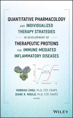
Bispecific antibodies (BsAbs) exhibit dual functionality and are capable of binding to two different epitopes of the same or different antigens. BsAbs can be divided into “IgG‐like” or “non‐IgG‐like” formats based on their resemblance to the native IgG. “IgG‐like” BsAbs have so far been the predominant group and are produced by a variety of methods, including the Quadroma [1], Knobs‐into‐Holes [2], CrossMab [3], dock‐and‐lock [4], DuoBody [5], and dual‐variable‐domain immunoglobulin [6] approaches. “Non‐IgG‐like” BsAbs have a lower molecular weight and include formats such as bispecific T‐cell engagers (BiTEs), dual‐affinity retargeting molecules (DARTs), and tandem diabody (TandAb) systems. In “non‐IgG‐like” BsAbs, antibody fragments, such as antibody‐binding fragment (Fab), single‐chain fragment variable (scFv), or single‐domain antibodies (sdAbs), are used as building blocks connected by short flexible peptide linkers. A recent review by Kontermann and Brinkmann exhibits the versatility of BsAb designs demonstrated by over 60 different formats that have been produced to date, with over 30 BsAbs in clinical development (Figure 9.1) [7].

Figure 9.1 A selection of different BsAbs scaffolds approved or under development. (See insert for color representation of this figure.)
Source: Kontermann and Brinkmann 2015 [7]. Reproduced with permission from Elsevier.
9.2 Quantitative Pharmacokinetic Considerations of BsAbs
BsAbs are known to have limited oral bioavailability due to low permeability through the gut wall and high gastrointestinal protease activity. Thus, parenteral routes of administration, particularly intravenous (IV) and subcutaneous (SC) administration, are the most frequently utilized dosing pathways. Direct delivery to the site of action has also been described for attaining high local concentrations [8].
Distribution of BsAbs is determined by molecular weight, physicochemical properties including charge, hydrophobicity, and binding to Fc receptors. A biexponential pharmacokinetic profile is typical for intravenously administered IgG like BsAbs, with a volume of distribution equal to or slightly larger than the plasma volume (3–8 l) representing the vascular space and interstitial space of well perfused organs [9–11]. The movement of BsAbs from the systemic circulation to the interstitial space is similar to other large proteins predominantly facilitated by convective transport rather than diffusion, thereby following the unidirectional fluid flux from the vascular space into the interstitial space. Subsequent removal from the interstitial space back into the vascular space is regulated by lymphatic drainage [12,13].
Clearance of larger IgG‐like BsAbs depends upon intracellular uptake and is usually facilitated by pinocytosis or similar endocytotic processes [10,14]. BsAbs are predominantly eliminated by catabolism resulting in peptides and amino acids that are reutilized for de novo protein synthesis. This nonspecific proteolytic degradation can be carried out ubiquitously throughout the body, predominantly by endothelial cells and the reticuloendothelial system [15,16]. In addition to this nonspecific clearance pathway, BsAbs may also undergo classical target‐mediated drug disposition after binding to one or both of their targets, where the target binding results in intracellular uptake and subsequent lysosomal degradation [17]. As this process can be saturated at therapeutic concentrations and may constitute a major elimination pathway for some BsAbs, nonlinear pharmacokinetic behavior with higher clearance and shorter half‐life at low concentrations and lower clearance and longer half‐life at higher concentrations is oftentimes observed.
Elimination via renal metabolism must be considered for smaller BsAbs (molecular weights less than 60 kDa), such as blinatumomab, as these can undergo glomerular filtration and subsequent catabolism by intracellular uptake into tubular cells and lysosomal degradation, resulting in potentially short elimination half‐lives [18].
The neonatal Fc receptor (FcRn) recycles IgG‐like BsAbs with intact Fc domain after intracellular uptake by preventing these molecules from lysosomal degradation [19]. Similar to endogenous IgG and monospecific monoclonal antibodies (mAbs), this salvage pathway also prolongs the elimination of IgG‐type BsAbs, while non‐IgG‐like BsAbs such as BiTes have in comparison substantially shorter half‐lives (blinatumomab: 1.25 ± 0.63 hours vs. emicizumab: 28–34 days) due to their lack of interaction with the FcRn salvage pathway due to the absence of a Fc domain with FcRn binding site [20,21].
9.3 Preclinical Considerations
9.3.1 Antibody Properties
It is well established that pharmacokinetic and pharmacodynamic properties of BsAbs are governed by both molecule‐dependent and species‐dependent factors [22]. Several biological processes are key determinants in antibody pharmacokinetics such as binding to antigens [17] and other cognate receptors [23]. Furthermore, a BsAb's valency, shape, size, isoelectric point (pI), and route of administration may also influence its disposition kinetics and clearance [24,25].
The charge on BsAbs can have a substantial impact on their disposition. Modifications of the isoelectric point of an antibody by one or more units can result in significant differences in its pharmacokinetics [26]. For example, an investigational IgG‐type BsAb for the treatment of hemophilia A, a precursor molecule to emicizumab, was found to have unexpectedly high clearance in mice. This was partially attributed to a large positive charge cluster in the variable region that might have increased the nonspecific binding to the extracellular matrix and subsequently increased its clearance. To overcome this limitation, a single Tyr30Glu mutation was carried out that markedly neutralized the charge cluster and increased the plasma half‐life without compromising its pharmacological activity [27].
9.3.2 Selection of a BsAb Format
With over 60 types of BsAb constructs explored to date [7], selection of a suitable BsAb construct is crucial for successful development and approval. The following criteria have been used to guide the selection process:
BsAbs with Fc region differ in their mechanism of action and pharmacokinetic behavior based on their molecular size. BsAbs with Fc region are larger in size, which bestows them longer elimination half‐life. The better pharmacokinetic profile is seen because the larger size does not allow them to undergo glomerular filtration and FcRn receptor binding helps in escaping lysosomal degradation [7]. BsAb derivatives without Fc region are prone to both elimination mechanisms: glomerular filtration with subsequent renal metabolism and lysosomal degradation. Another advantage of the presence of the Fc region is the ability to elicit immune system‐specific effector mechanisms such as antibody‐dependent cellular cytotoxicity (ADCC), complement‐dependent cytotoxicity (CDC), and antibody‐dependent cellular phagocytosis (ADCP) for destroying target cells [28]. An IgG‐like BsAb, catumaxomab is the only BsAb approved in this category containing epitopes specific for binding to malignant cells and T cells, as well as one arm (Fc region) dedicated to recruit accessory cells like natural killer cells, macrophages, or dendritic cells to the target site [29].
BsAb constructs without Fc region are relatively smaller size BsAbs. Antigen recognizing sites (epitopes) are bridged with short amino acid chains that impart flexibility and close association of target and effector cells. BiTEs are so far the most successful construct design in this category, and blinatumomab [30] is the only BsAb approved in this category to date. BiTes are generated by connecting two single chain variable domains with a short flexible linker. For blinatumomab, one arm is specific for the CD3 receptors on T cells, and the other arm is engineered to bind with malignant cell surface receptors [31,32]. The major disadvantage of BsAbs without Fc region is rapid elimination from the body and hence, dosing regimens with either frequent discrete or continuous parenteral administration are warranted for desired exposure to achieve the intended pharmacological effects.
9.3.3 In vitro Models
For blinatumomab, in vitro target‐binding assays were performed on CD3‐expressing Jurkat cells and CD19‐expressing cell lines including Blin‐1 (recapitulates the pre‐B to B‐cell stage transition) and Daudi (a B‐lymphoblast cell line) [33,34]. Blinatumomab did not show binding in cell lines lacking expression of CD19 or CD3, suggesting the selectivity toward CD19 and CD3 receptors. Tumor cell lysis activity was tested in target cell lines co‐expressing CD19 and CD20 (Raji, Karpas‐422, and ARH‐77) and human peripheral blood mononuclear cells (PBMCs, a source of T cells) as effectors. Blinatumomab achieved 60–100% target cell lysis in a 24‐hour incubation period [35]. Lysis of CD19 expressing B cells observed in in vitro studies suggests that the mechanism of cell lysis is not driven by CDC or ADCC but rather a cell‐mediated mechanism in which toxic proteins (perforin–granzyme) are being released upon formation of a cytolytic synapse between T cells and target cells. These studies have also been conducted on T cells derived from healthy donors cocultured with CD19+ target cells. The blinatumomab EC50 obtained in these assays was in the range of 10–100 pg ml−1 [36]. Blinatumomab was also tested in homologous settings where target cells from non‐Hodgkin lymphoma (NHL) or acute lymphocytic leukemia (ALL) cell lines were investigated for their activity against PBMC samples from chronic lymphocytic leukemia (CLL) patients. The results from these in vitro studies confirm that blinatumomab had excellent activity against B cells and was a good drug candidate to perform in vivo efficacy studies.
With larger IgG‐type BsAbs, such as emicizumab, in vitro cross‐reactivity in cynomolgus monkeys was assessed by an activated partial thromboplastin time (APTT) assay. This evaluation showed that emicizumab shortened APTT in factor VIII (FVIII)‐neutralized cynomolgus monkey plasma in a concentration‐dependent manner similar to human FVIII‐deficient plasma. Furthermore, by utilizing a thrombin generation (TG) assay, it was also demonstrated that emicizumab improved the response of FXIa‐triggered TG assay in FVIII‐neutralized cynomolgus monkey plasma in a concentration‐dependent manner similar to human plasma. These in vitro evaluations demonstrated that emicizumab had a similar cofactor activity as rhFVIII or rpoFVIII in improving the peak height in each species.
9.3.4 In vivo Models
Several parameters must be considered prior to evaluation of BsAbs in animal models, including antibody cross‐reactivity, immunogenicity, pharmacokinetics, and possible interactions with the host immune system. A few considerations were suggested and previously reviewed by Loisel et al. [37] and are listed below:
- Does the BsAb directed to a human target recognize the animal antigen, and on which type of cells? Consequently, is it necessary to “implant” the human antigen/target cell in the chosen animal model?
- Does the BsAb raise an immunogenic response in the animal model, and if so, how will the induced anti‐drug antibodies (ADAs) affect the results?
- Can the BsAb half‐life and the biodistribution be extrapolated to the human situation, and what are the identified causes of possible differences (e.g. soluble receptors, FcRn)?
- How does the BsAb interfere with the animal immune system (e.g. ADCC, complement activation, targeting of NK cells)?
Selection of an appropriate rodent efficacy model is critical to BsAb discovery, development, and translational strategies. For anti‐cancer drugs, the transgenic knock‐in mouse model is an informative tool to evaluate antibodies that exhibit poor or no cross‐reactivity to the murine ortholog of the antigen target [38,39]. Moreover, these models allow to circumvent the use of surrogate antibodies and the associated investments and production challenges. Use of orthotopic models allow implantation of human tumors to mimic clinical‐like tumor growth and metastasis [40]. Also, primary tumor fragments, as opposed to cell lines, are histologically intact, maintain tissue architecture, and preserve and reflect the original genetic lesions of the disease [41].
Another major hurdle in the early clinical investigation of BsAbs is the high inherent risk of adverse drug reactions in humans [42]. Risk prediction of such adverse reactions and dose selection for first‐in‐human (FIH) studies are based on preclinical safety assessments in at least one pharmacologically relevant animal species [43]. Selection of a relevant animal model and data interpretation of toxicity studies in such a model are paramount. Owing to the similar developmental challenges for mAbs and BsAbs much of the development strategies for BsAbs are based on current science with mAbs. Due to genetic and pharmacological similarity to humans, nonhuman primates (NHPs) are the most commonly selected animal models for the safety assessment of BsAbs. In the absence of pharmacologically relevant animal models, use of a surrogate mouse BsAb (recognizing the mouse homolog of the human target) [44] or a human BsAb in human target transgenic mice may be potential alternatives, which are dependent on the specific pharmacological/toxicological endpoints intended to be assessed and whether they are likely to be predictive of the outcome in humans in these alternative toxicology models.
9.3.5 Catumaxomab
The trifunctional antibody (trAb) catumaxomab targets epithelial cell adhesion molecule (EpCAM) on tumor cells, CD3 on T cells, and the Fcγ‐receptor of antigen‐presenting cells. Due to the specificity of catumaxomab for its human target antigens, a targeted preclinical testing strategy was developed in standard animal models. This strategy did not result in abnormal or test substance‐related acute toxicity or local intolerance at the administration site [45]. The antigenicity and immunotoxicity of catumaxomab were investigated in the cynomolgus monkey. No immunotoxic effects were observed following IV infusion of catumaxomab, and there was no effect on circulating levels of cytokines or complement. The detection of ADAs in the monkey was expected since catumaxomab is a mouse/rat BsAb that is recognized as a foreign (immunogenic) molecule by the animal's immune system.
The in vivo anti‐tumor activity of catumaxomab was evaluated in an immunologically compromised murine model of ovarian carcinoma using the variant antibody BiLu (anti‐mouse CD3 × anti‐human EpCAM). BiLu has a structure equivalent to catumaxomab but binds to mouse CD3. BiLu was highly active resulting in 100% survival (control group had no survivors beyond 28 days). Additionally, a long‐lasting antitumor immunity was observed in 14 out of 18 mice [46].
In a preclinical pharmacokinetic study, catumaxomab was administered to female C.B‐17 severe combined immunodeficiency (SCID) mice as either IV (Group I) or intraperitoneal (IP) (Groups II–IV) injection at 100 µg kg−1. To evaluate the influence of tumor load and immune effector cell numbers at the site of application on the influence the pharmacokinetics, two additional groups of mice, Groups III and IV received SKOV‐3 tumor (American type culture collection, ATCC HTB‐77) and PBMC effector cells, both at 2 × 10⁶ (Group III) or 1 × 10⁷ (Group IV) per mouse were included. Pharmacokinetic parameters were based on the mean plasma concentrations of catumaxomab. In the absence of binding partners, the observed IP bioavailability of catumaxomab was 82%. The bioavailability significantly declined to 68% and 27% in the presence of low or high levels, respectively, of tumor and effector cells [47].
9.3.6 Emicizumab
Emicizumab (ACE910) is a humanized anti‐FIXa/FX bispecific IgG antibody mimicking the cofactor function of FVIII and was developed to treat hemophilia A. In vivo hemostatic potency of emicizumab was evaluated in a cynomolgus monkey model with acquired hemophilia A. After injury, emicizumab when administered at 1 and 3 mg kg−1 lead to a significant reduction of bruised areas (P < 0.05 vs. control) [48].
In a preclinical pharmacokinetic study, cynomolgus monkeys were treated with either a single IV (6 mg kg−1; n = 2) or SC (0.06, 0.6, or 6 mg kg−1; n = 3) dose. The plasma half‐life of emicizumab was 19.4 days after a single IV administration at 6 mg kg−1 and was 23.6–26.5 days after a single SC administration at 0.06, 0.6, or 6 mg kg−1, with a 102.3% bioavailability. From the in vivo hemostatic studies in NHPs, the average initial plasma concentrations of emicizumab were either 26 or 61 µg ml−1 after administration of 1 or 3 mg kg−1 doses, respectively. Hemostatic effects observed in these groups were comparable to rpoFVIII 10 U kg−1 group, in which the FVIII level was within the range of a mild phenotype. To convert a severe phenotype to a mild phenotype, by routine supplementation, a plasma emicizumab level higher than 26 µg ml−1 was needed in patients. Hence, multiple‐dosing simulations of emicizumab were explored. The results of the simulations suggested that the target trough plasma levels of emicizumab at 26 or 61 µg ml−1 could be maintained by a once‐weekly SC administration of 0.64 or 1.5 mg kg−1 at steady state, respectively. This formed the basis for the dosing schedule for the first phase I clinical trial [49].
9.3.7 Blinatumomab
Blinatumomab is the first BiTE, approved for treatment of ALL. Due to its small molecular size (54.1 kDa), blinatumomab facilitates bridging cytotoxic T cells with CD19 positive lymphoma cells. One of the epitopes of this BsAb engages in recruiting T cells and redirecting them toward cancerous cells with the help of the selectivity of another epitope [29,50]. Blinatumomab demonstrates a stronger affinity toward CD19 (1.49 × 10⁻⁹ M) than CD3 receptors (2.6 × 10⁻⁷ M) [36,51]. The mechanism of action indicates that blinatumomab does not need CD3 specificity, and hence, it is associated with polyclonal T‐cell activation.
Preclinical species selection for in vivo evaluation of blinatumomab was based on binding to CD3 and CD19 receptors on PBMCs. These results demonstrated that chimpanzees are the most relevant nonhuman species. Being an endangered species, ethical concerns diverted further development activities toward mouse models [52]. A surrogate BiTE (muS103new) specific for murine CD19 and CD3 was engineered to perform further safety and efficacy studies in mouse models [53]. Non‐obese diabetic (NOD)/SCID mice xenograft models were used to evaluate anti‐tumor activity. Of the eight studies reported with either IV or SC administration in mice implanted with NALM‐6, SEMc, Raji, and Granta tumors, blinatumomab showed statistically significant inhibition of tumor growth and prolongation of median survival at doses above 1 µg d−1 [53]. For pharmacokinetic studies in mice, blinatumomab demonstrated a dose‐proportional increase in exposure after IV administration and had an SC bioavailability of 35%. Rat and monkey studies with blinatumomab had longer half‐lives with SC administration ranging from five to eight hours. In chimpanzees, blinatumomab was administered by a two‐hour IV infusion once weekly, with half‐lives ranging from 1.5 to 2.6 hours.
9.3.8 Anti TfR/BACE1
Delivery of protein therapeutics to the brain is challenging [54,55] since several barriers like the blood–brain‐barrier (BBB), the blood‐cerebrospinal fluid barrier (BCSFB), and the meningeal layers limit drug absorption to the brain parenchyma. Despite limited permeability of large molecules such as antibodies across the brain, it was previously demonstrated that proteins can successfully be transported across the BBB [54], but concentrations remain limited due to poor penetration into the brain (∼0.1% of serum concenrtation was available in brain). A novel approach of transporter‐mediated transcytosis to cross the BBB was used in the development of anti‐TfR/BACE1 (transferrin receptor and β‐secretase 1). The transferrin receptor (TfR) is a transcytotic receptor expressed on endothelial cells, including those of the BBB. BACE1 is an enzyme responsible for the initial cleavage of amyloid precursor protein (APP), which is further cleaved by gamma‐secretase, giving rise to amyloid‐β peptides (Aβ40 and Aβ42), the primary components of amyloid plaques found in the brains of Alzheimer's disease patients [56,57]. Here, a BsAb is targeting with one arm a transporter at the BBB and the other arm is designed to bind to targets in the brain to treat neurological disorders. Anti‐TfR/BACE1 uses the iron transporter protein transferrin to enter the brain and then bind to and inhibit the BACE1 [58–60].
Studies by Couch et al. investigated targeting TfR to achieve brain delivery of therapeutic antibodies [61]. Studies conducted in rodents with several affinity variants of BsAb against mouse TfR were evaluated for pharmacokinetic and pharmacodynamic properties. The lower affinity variants could not reach the brain due to insufficient interaction with TfR transporters at BBB epithelial cells, whereas very high affinity variants are retained in the brain epithelium and cleared by lysosomal degradation before entering brain tissue. Titration of TfR affinity by balancing TfR binding at the BBB and release into the brain was a pivotal exercise in the development of the anti‐TfR/BACE1 BsAb. Another goal of the study was to assess the safety of anti‐TfR/BACE1. In addition to epithelial cells at the BBB, immature red blood cells (reticulocytes) are also rich in TfR expression. The results suggested that anti‐TfR/BACE1 is liable for reticulocyte loss associated with anti‐TfR function.
Further screening of anti‐TfR/BACE1 was performed in primates and a human TfR knock‐in mouse model by Yu et al. [58]. Two humanized BsAbs, anti‐TfR1/BACE1 (high TfR affinity) and anti‐TfR2/BACE1 (low TfR affinity) were generated using “knob in hole” technology. A brain uptake study conducted after dosing 50 mg kg−1 in mice showed higher plasma and brain concentrations for the low‐affinity variant (anti‐TfR2/BACE1) compared to the high‐affinity variant (anti‐TfR1/BACE1). However, the extent of β‐amyloid reduction as a pharmacodynamic biomarker was similar for both BsAbs, suggesting that antibody concentrations exceeded the in vivo concentrations needed to drive β‐amyloid reduction. After administration of 30 mg kg−1 to cynomolgus monkeys, both these variants yielded conflicting results relative to the human TfR knock‐in mouse. TfR1/BACE1 (a high‐affinity variant) showed higher and more sustained systemic exposure in brain and plasma, and also a more pronounced and sustained reduction in β‐amyloid than TfR2/BACE1. As these BsAbs were designed against human TfR, the low‐affinity variant, TfR2/BACE1 had relatively much lower affinity toward monkey TfR to adequately interact and be transported into the brain by transcytosis. Toxicity studies after dosing 30 mg kg−1 of both low‐ and high‐affinity variants in monkey demonstrated no sign of reticulocyte loss and associated symptoms [58].
9.4 Translational Considerations
Many mAbs employed in cancer therapy mediate at least part of their anti‐tumor activity by ADCC and/or CDC. The therapeutic activity of these mAbs is limited by a relatively low affinity against Fcγ receptors on effector cells and high competition with abundant endogenous IgG [31,62]. T‐cell recruiting BsAbs have an advantage over traditional mAbs as one of their arms engages in recruiting immune cells to the target, the cancer cell, identified by the second arm of the BsAb.
After initial preclinical evaluation, successful lead BsAbs with desirable safety and efficacy properties are further evaluated in clinical studies. The vital task prior to clinical studies is the estimation of a safe and practical FIH dose. There have repeatedly been situations where an animal model completely failed to predict the toxicity in humans. For instance, in a phase I clinical trial with TGN1412 [63], a recombinantly expressed, humanized superagonist anti‐CD28 mAb, a starting dose of 1.5 µg kg−1 caused life‐threatening cytokine release syndrome in healthy volunteers [63,64]. The reason for this was failure to identify a relevant animal model on which dose extrapolations could be based. This interpretation was further proven by administering a range of doses (0.1, 0.5, 5, or 50 mg kg−1) to cynomolgus monkeys that did not experience any adverse reactions in contrast to humans [65]. The underlying basis for this adverse reaction was later established by Eastwood et al. In cynomolgus monkeys, T cells that develop into CD4+ effector memory T cells lose their CD28 receptors. Thus, these cells would not be activated by TGN1412. In humans, however, CD4+ effector memory T cells retain CD28 receptors on their surface. Thus, human CD4+ effector memory T cells are still activated by TGN1412 and therefore rapidly produce pro‐inflammatory cytokines [66].
TGN1412 was subsequently further developed by another company under the name TAB08. In a new study, an extremely low starting dose was chosen, 0.1% of the dose used in the initial FIH study (and therefore 0.0002% of the maximum dose used in the original cynomolgus monkey study). At this new starting dose, only 1% of human CD28 receptors would be bound by TGN1412/TAB08 antibodies. During the clinical study, the dose was increased by small increments in different cohorts, and all subjects were monitored closely. No serious side effects were observed [67].
Several approaches are concurrently used to estimate FIH including safety‐based methods such as maximum recommended starting dose (MRSD) and human equivalent dose (HED) for safety, as well as pharmacology‐based methods such as minimum anticipated biological effect level (MABEL), and human pharmacologically active dose (hPAD) [64].
The FIH dose level should be high enough to show some, but very limited pharmacological activity and low enough not to show any adverse effects. An already complex task becomes more challenging while dealing with antibody therapies that mediate T cell response. Therapeutic mAbs that are potent T cell agonists have a high potential to elicit immune hypersensitivity reactions. In such cases, the ICH S9 guideline recommends use of pharmacology‐driven MABEL approach for the selection of a starting dose in humans [68]. MABEL is the minimum dose/concentration required to produce intended pharmacological activity. The conventional MABEL approach is based on the estimation of the dose/concentration of the therapeutically active moiety, where a single moiety is responsible for producing pharmacological activity. In case of BsAbs recruiting immune cells to kill cancer cells, the use of BsAb concentration alone is not appropriate for the estimation of MABEL. A tri‐molecular synapse assembly (BsAbs, immune cells, and cancer cells) triggers a pharmacological response and hence concentration of the tri‐molecular synapse instead of the BsAb concentration is more appropriate for the estimation of MABEL in case of these BsAbs. Chen et al. used, for example a pharmacokinetic/pharmacodynamic‐driven approach for projecting MABEL of P‐cadherin LP‐DART [69].
9.5 Immunogenicity
Most BsAbs elicit some level of antigenicity, leading to endogenous antibody response (ADAs) against the therapeutic product when administered to humans. These ADAs that bind to the therapeutic protein may have either a neutralizing or nonneutralization effect with regard to target interaction. Even the nonneutralizing ADAs may reduce the therapeutic activity by triggering an additional clearance pathway for the therapeutic protein through immune complex formation and subsequent degradation [70]. Compared to the monospecific mAbs and IgG‐type BsAbs, single‐chain variable fragment‐based platforms such as tandem scFv and diabodies are generally less immunogenic due to the absence of an Fc domain in the molecule [71,72]. Bispecific diabodies are even less likely to prompt immune reactions due to their compact size [73]. Thus, small bispecific constructs may have a substantial clinical advantage compared to classic mAbs with regard to immunogenic potential.
9.6 Clinical Development of BsAbs
9.6.1 Catumaxomab
Catumaxomab is a trAb indicated for the IP treatment of malignant ascites [74] in patients with EpCAM‐positive [75–77] epithelial cell cancers such as ovarian, gastric, lung, breast, colon, and prostate cancer [78–82], where standard therapy is not available or no longer effective. Catumaxomab resulted in a significant increase in survival of 10–24 months in patients with ovarian cancer, followed by patients with breast cancer (1–6 months), and GI cancers (1–3 months) [81,83].
In a multicenter phase II study to determine local and systemic antibody concentrations and ADA development, 13 patients (primary tumor types: ovarian (69%), pancreatic (23%), and gastric (8%) carcinoma) with symptomatic malignant ascites were treated with four ascending doses of 10, 20, 50, and 150 µg catumaxomab by IP infusion on Days 0, 3, 6, 7 and 10. Catumaxomab was concentrated in the ascites fluid, attaining immunologically active concentrations in circulation after several days of the IP infusion. Systemic catumaxomab exposures were low (<1% bioavailability), with a maximal median plasma concentration of 403 pg ml−1 and a mean plasma elimination half‐life of 2.13 days. All patients developed ADA after the last infusion, i.e. within 11–16 days [84]. High inter‐individual variability and low systemic exposure may be explained by the inverse correlation between tumor burden, effector cell numbers, and systemic antibody bioavailability as demonstrated in a defined mouse tumor model [47].
In a phase II/III trial [84], cancer patients (n = 258) with recurrent symptomatic malignant ascites, resistant to conventional chemotherapy were randomized to paracentesis plus catumaxomab (catumaxomab) or paracentesis alone (control) and stratified by cancer type (129 ovarian and 129 nonovarian). Catumaxomab was administered as four 6‐hour constant‐rate IP infusions on Days 0, 3, 7, and 10 at doses of 10, 20, 50, and 150 µg. Patients were assessed in up to five follow‐up visits at eight days, 1, 3, 5, and 7 months (end of study) after the last infusion (catumaxomab group) or therapeutic paracentesis (Day 0, control group). In the control group, the study reached an end when the patient required the next paracentesis or died, whichever occurred first. The primary efficacy endpoint was puncture‐free survival as the patients with symptomatic malignant ascites have a very short life expectancy (few weeks to months). Secondary efficacy parameters included time for next paracentesis, ascites signs and symptoms, and overall survival. Puncture‐free survival was significantly longer in the catumaxomab group (median 46 days) than the control group (median 11 days) (hazard ratio = 0.254; P < 0.0001) as was median time to next paracentesis (77 vs. 13 days; P < 0.0001). In addition, catumaxomab patients had fewer signs and symptoms of ascites than control patients. In a prospectively planned analysis, overall survival for the catumaxomab group and was significantly prolonged in patients with gastric cancer (n = 66; 71 vs. 44 days; P = 0.0313). The next therapeutic puncture in the control group (13 days) compared to the catumaxomab group (77 days) indicated that catumaxomab treatment avoided the need for approximately five punctures. These results demonstrated that paracentesis plus catumaxomab had superior efficacy compared to paracentesis alone for the treatment of malignant ascites. A subgroup analysis of gastric cancer patients, the largest subpopulation in the nonovarian stratum, demonstrated a statistically significant prolongation of overall survival.
9.6.2 Emicizumab
The primary objective of emicizumab is to abate the problems caused by prophylactic IV infusions of FVIII, which has a substantially shorter in vivo half‐life than emicizumab [85]. In a FIH inter‐individual dose‐escalation study [86], healthy Japanese and white male subjects (aged 20–44 years) were grouped based on their ethnicity. Japanese subjects were randomized to receive a single SC injection of emicizumab (0.001, 0.01, 0.1, 0.3, or 1 mg kg−1; n = 6 per dose group) or placebo (n = 2 per dose group). White subjects were randomized to receive a single SC injection of emicizumab (0.1, 0.3, or 1 mg kg−1; n = 6 per dose group) or placebo (n = 2 per dose group). Subjects were either observed for 4 weeks (0.001 or 0.01 mg kg−1), 16 weeks (0.1 mg kg−1), 20 weeks (0.3 mg kg−1), or 24 weeks (1 mg kg−1). Pharmacokinetic parameters were calculated for doses of 0.01 mg kg−1 and higher. Emicizumab showed linear dose–exposure profiles in both groups, Japanese and white subjects, with Cmax ranging from 0.0675 to 5.92 mg ml−1 (0.01–1 mg kg−1) and AUC ranging from 30.2 to 304 µg d ml−1 (0.1–1 mg kg−1). The half‐life of emicizumab was 28.3–34.4 days, similar to the half‐life observed in cynomolgus monkeys (23.6–26.5 days at doses of 0.06, 0.6, and 6 mg kg−1) [49]. The pharmacokinetic and safety profiles of emicizumab were found to be similar between Japanese and white healthy subjects, consistent with observations for other antibody‐based drugs [87]. Generally, racial differences in body weight and target antigen levels may contribute to the pharmacokinetic profile of antibody drugs [22]. With its longer half‐life compared to FVIII, it was concluded that emicizumab is expected to be a more effective prophylactic treatment of hemophilia with potential for a more convenient once‐weekly, SC injection regimen.
In another dose‐escalation study of emicizumab, 18 Japanese patients with severe hemophilia A were divided equally into three cohorts: patients received SC emicizumab (80 mg ml−1) at an initial dose of 1 mg kg−1 of body weight (Cohort 1) or 3 mg kg−1 (Cohorts 2 and 3) at Week 0 (Day 1), followed by a once weekly SC dose of 0.3, 1, or 3 mg kg−1 (Cohorts 1, 2, and 3 respectively) from Week 1 to Week 12. Upon treatment with emicizumab, the bleeding rate was reduced markedly from the rate at baseline in all cohorts. The annualized bleeding rate decreased among 17 patients regardless of the presence or absence of FVIII inhibitors and prior prophylaxis.
9.6.3 Blinatumomab
Blinatumomab has two single chain variable fragments specific for the CD3 and CD19 receptors connected by a five nonimmunogenic amino acids linker. The lack of Fc region in conjunction with its low molecular weight (below the glomerular filtration cutoff) results in rapid clearance of blinatumomab. In contrast to the studies in preclinical species, this rapid elimination did not allow discrete IV dosing in clinical studies but required continuous infusion to maintain therapeutic plasma concentration for a sufficient period of time in order to achieve the desired pharmacological activity [88,89]. The first clinical study was conducted by administrating blinatumomab by short‐term IV administration (two to four hours), but all subsequent studies were performed with continuous intravenous infusion for one week. Six phase I and II studies have been conducted on blinatumomab for the safety and efficacy evaluation. In the six clinical studies, blinatumomab exhibited linear pharmacokinetics when evaluated over a dose range of 5–90 µg m−2 d−1, administered as continuous infusions up to eight weeks to patients (n = 499) with acute lymphoblastic leukemia and non‐Hodgkin's lymphoma. Typical pharmacokinetic parameters obtained by noncompartmental analysis from these studies were a clearance of 2.72 ± 2.71 l h−1, a volume of distribution of 4.52 ± 2.89 l and a half‐life of 2.11 ± 1.42 hours [30]. The pharmacokinetic parameters were independent of gender, age, and disease type in the populations assessed. These studies formed the basis for dose selection for a main phase II, open‐label, single‐arm, multicenter trial to evaluate efficacy and safety in adults with relapsed/refractory B‐precursor acute lymphoblastic leukemia. All subjects (n = 189) received one to five cycles of blinatumomab as a continuous IV infusion at an initial dose of 9 µg d−1 for the first seven days of cycle 1. The median OS for all subjects was 6.1 months. The median observation time was 9.8 months; 6‐ and 12‐month survival probabilities were 50%. Overall, blinatumomab demonstrated antitumor activity in phase I–II trials in patients with NHL, minimal residual disease‐positive ALL (MRD+ ALL), and r/r ALL. Blinatumomab was granted breakthrough therapy designation by the US Food and Drug Administration in July 2014 and approved by the end of that year.
9.7 Conclusion
BsAbs are capable of simultaneously acting on two different pharmacological targets that is an attractive feature and can aide in serving as a lucrative bridge between therapeutics and targets. The development of BsAbs offers promising new therapeutic strategies that may ultimately translate into improved treatment modalities in a variety of indications. Numerous preclinical and clinical development projects are currently ongoing to move BsAbs closer to this overall goal.
While some challenges during the development process are similar to other therapeutic proteins, many new challenges need to be addressed. Limited availability of in vitro models and their lack of potential translational capability to predicting activity in the selection of lead candidates for trials present a significant challenge. Although, several in vivo disease‐specific models such as transgenic knock‐in mouse models are available for screening, their utilization and predictive value often suffer from lack of cross‐reactivity and/or immunogenicity. In some cases, the most relevant in vivo model being an endangered species (chimpanzees for blinatumomab) presents a challenge for translation. Surrogate molecules utilized for development should be treated with caution in predicting a FIH dose as previously noted in the case of development of TGN1412. Penetrating biological membranes to achieve desired concentrations in peripheral target organs like the brain has always been a problem with large molecules but use of endogenous transport systems has been explored to overcome that issue.
Despite several successful development programs, a more rapid progress in learning and understanding the key parameters for the development success of BsAbs, however, seems at the current time to be hampered by a variety of factors, including the following:
- There is a multitude of BsAbs scaffolds available and possible, and identifying a preferred scaffold is a complex process.
- Many scaffolds have intellectual property space build around them that necessitates companies to explore even more new constructs.
- Due to the large number of scaffolds, learning crucial aspects of each scaffold is a new learning experience and thus overall knowledge gain in development is fragmented and overall development progress is slow.
Although, the majority of BsAbs is developed for treatment of various cancers, several new types of BsAbs are now also being developed for other indications such as rheumatoid arthritis, for example ozaralizumab [90], treatment of age‐related macular degeneration [91,92], idiopathic pulmonary fibrosis [93], Alzheimer's disease [94], and HIV‐infection [95]. Smaller BsAbs such as BiTes that may have significant advantages such as ease of manufacturing and enhanced tissue penetration suffer from short half‐lives due to their lack of the Fc region, while larger IgG such as BsAbs can circulate in the plasma for days and weeks. Strategies such as PEGylation [96], fusion with human serum albumin [97], multimerization [96], or Fc‐fragment fusion [98] may aid significantly in prolonging the half‐life of bispecific constructs.
Overall, BsAbs offer an attractive platform technology to further harness the promise of precision medicine by creating targeted and highly specific therapeutics for unmet medical needs that improve the risk–benefit ratio in many difficult‐to‐treat indications.
References
- 1 Lindhofer, H., Mocikat, R., Steipe, B., and Thierfelder, S. (1995). Preferential species‐restricted heavy/light chain pairing in rat/mouse quadromas: implications for a single‐step purification of bispecific antibodies. J. Immunol. 155 (1): 219–225.
- 2 Spiess, C., Merchant, M., Huang, A. et al. (2013). Bispecific antibodies with natural architecture produced by co‐culture of bacteria expressing two distinct half‐antibodies. Nat. Biotechnol. 31 (8): 753–758.
- 3 Schaefer, W., Regula, J.T., Bähner, M. et al. (2011). Immunoglobulin domain crossover as a generic approach for the production of bispecific IgG antibodies. Proc. Natl. Acad. Sci. 108 (27): 11187–11192.
- 4 Dmitrieva, N.I., Cui, K., Kitchaev, D.A. et al. (2011). DNA double‐strand breaks induced by high NaCl occur predominantly in gene deserts. Proc. Natl. Acad. Sci. 108 (51): 20796–20801.
- 5 Schuurman, J., Graus, Y.F., Labrijn, A.F. et al. (2014). Opening the door to innovation. MAbs 6 (4): 812–819.
- 6 Wu, C., Ying, H., Grinnell, C. et al. (2007). Simultaneous targeting of multiple disease mediators by a dual‐variable‐domain immunoglobulin. Nat. Biotechnol. 25 (11): 1290–1297.
- 7 Kontermann, R.E. and Brinkmann, U. (2015). Bispecific antibodies. Drug Discovery Today 20 (7): 838–847.
- 8 Rathi, C. and Meibohm, B. (2015). Clinical pharmacology of bispecific antibody constructs. J. Clin. Pharmacol. 55 (Suppl. 3): S21–S28.
- 9 Portell, C.A., Wenzell, C.M., and Advani, A.S. (2013). Clinical and pharmacologic aspects of blinatumomab in the treatment of B‐cell acute lymphoblastic leukemia. Clin. Pharmacol. 5 (Suppl. 1): 5–11.
- 10 Tang, L., Persky, A.M., Hochhaus, G., and Meibohm, B. (2004). Pharmacokinetic aspects of biotechnology products. J. Pharm. Sci. 93 (9): 2184–2204.
- 11 Ryman, J.T. and Meibohm, B. (2017). Pharmacokinetics of monoclonal antibodies. CPT Pharmacometrics Syst. Pharmacol. 6 (9): 576–588.
- 12 Rippe, B. and Haraldsson, B. (1994). Transport of macromolecules across microvascular walls: the two‐pore theory. Physiol. Rev. 74 (1): 163–219.
- 13 Meibohm, B. (2012). Pharmacokinetics and half‐life of protein therapeutics. In: Therapeutic Proteins: Strategies to Modulate Their Plasma Half‐lives, vol. 48 (ed. R. Konterman).
- 14 Lobo, E.D., Hansen, R.J., and Balthasar, J.P. (2004). Antibody pharmacokinetics and pharmacodynamics. J. Pharm. Sci. 93 (11): 2645–2668.
- 15 Tang, L. and Meibohm, B. (2006). Pharmacokinetics of peptides and proteins. In: Pharmacokinetics and Pharmacodynamics of Biotech Drugs (ed. B. Meibohm). Weinheim: Wiley‐VCH.
- 16 Tabrizi, M.A., Tseng, C.‐M.L., and Roskos, L.K. (2006). Elimination mechanisms of therapeutic monoclonal antibodies. Drug Discovery Today 11 (1–2): 81–88.
- 17 Mager, D.E. (2006). Target‐mediated drug disposition and dynamics. Biochem. Pharmacol. 72 (1): 1–10.
- 18 Meibohm, B. and Zhou, H. (2012). Characterizing the impact of renal impairment on the clinical pharmacology of biologics. J. Clin. Pharmacol. 52 (S1): 54S–62S.
- 19 Roopenian, D.C. and Akilesh, S. (2007). FcRn: the neonatal Fc receptor comes of age. Nat. Rev. Immunol. 7 (9): 715–725.
- 20 Dirks, N.L. and Meibohm, B. (2010). Population pharmacokinetics of therapeutic monoclonal antibodies. Clin. Pharmacokinet. 49 (10): 633–659.
- 21 Diao, L. and Meibohm, B. (2013). Pharmacokinetics and pharmacokinetic–pharmacodynamic correlations of therapeutic peptides. Clin. Pharmacokinet. 52 (10): 855–868.
- 22 Wang, W., Wang, E.Q., and Balthasar, J.P. (2008). Monoclonal antibody pharmacokinetics and pharmacodynamics. Clin. Pharmacol. Ther. 84: 548–558.
- 23 Yeung, Y.A., Leabman, M.K., Marvin, J.S. et al. (2009). Engineering human IgG1 affinity to human neonatal Fc receptor:impact of affinity improvement on pharmacokinetics in primates. J. Immunol. 182: 7663–7671.
- 24 Beckman, R.A., Weiner, L.M., and Davis, H.M. (2007). Antibody constructs in cancer therapy: protein engineering strategies to improve exposure in solid tumors. Cancer 109: 170–179.
- 25 Boswell, C.A., Deng, R., Lin, K. et al. (2009). In vitro in vivo correlations of pharmacokinetics, pharmacodynamics and metabolism for antibody therapeutics. In: Proteins and Peptides: Pharmacokinetic, Pharmacodynamic, and Metabolic Outcomes (ed. R.J. Mrsny and A. Daugherty). New York: Informa HealthCare.
- 26 Boswell, C.A., Tesar, D.B., Mukhyala, K. et al. (2010). Effects of charge on antibody tissue distribution and pharmacokinetics. Bioconjugate Chem. 21 (12): 2153–2163.
- 27 Sampei, Z., Igawa, T., Soeda, T. et al. (2013). Identification and multidimensional optimization of an asymmetric bispecific IgG antibody mimicking the function of factor VIII cofactor activity. PLoS One 8 (2): e57479.
- 28 Holliger, P., Wing, M., Pound, J.D. et al. (1997). Retargeting serum immunoglobulin with bispecific diabodies. Nat. Biotechnol. 15 (7): 632–636.
- 29 Huehls, A.M., Coupet, T.A., and Sentman, C.L. (2015). Bispecific T‐cell engagers for cancer immunotherapy. Immunol. Cell Biol. 93 (3): 290–296.
- 30 Zhu, M., Wu, B., Brandl, C. et al. (2016). Blinatumomab, a bispecific T‐cell engager (BiTE®) for CD‐19 targeted cancer immunotherapy: clinical pharmacology and its implications. Clin. Pharmacokinet. 55 (10): 1271–1288.
- 31 Chames, P., Van Regenmortel, M., Weiss, E., and Baty, D. (2009). Therapeutic antibodies: successes, limitations and hopes for the future. Br. J. Pharmacol. 157 (2): 220–233.
- 32 Baeuerle, P.A., Kufer, P., and Bargou, R. (2009). BiTE: teaching antibodies to engage T‐cells for cancer therapy. Curr. Opin. Mol. Ther. 11 (1): 22–30.
- 33 Yao, R., Rich, S.A., and Schneider, E. (2002). Validation of sixteen leukemia and lymphoma cell lines as controls for molecular gene rearrangement assays. Clin. Chem. 48 (8): 1344–1351.
- 34 Wörmann, B., Anderson, J.M., Liberty, J.A. et al. (1989). Establishment of a leukemic cell model for studying human pre‐B to B cell differentiation. J. Immunol. 142 (1): 110–117.
- 35 d'Argouges, S., Wissing, S., Brandl, C. et al. (2009). Combination of rituximab with blinatumomab (MT103/MEDI‐538), a T cell‐engaging CD19‐/CD3‐bispecific antibody, for highly efficient lysis of human B lymphoma cells. Leuk. Res. 33 (3): 465–473.
- 36 Bumma, N., Papadantonakis, N., and Advani, A.S. (2015). Structure, development, preclinical and clinical efficacy of blinatumomab in acute lymphoblastic leukemia. Future Oncol. 11 (12): 1729–1739.
- 37 Loisel, S., Ohresser, M., Pallardy, M. et al. (2007). Relevance, advantages and limitations of animal models used in the development of monoclonal antibodies for cancer treatment. Crit. Rev. Oncol./Hematol. 62 (1): 34–42.
- 38 Bugelskil, P., Herzykl, D., Rehm, S. et al. (2000). Preclinical development of keliximab, a Primatized™ anti‐CD4 monoclonal antibody, in human CD4 transgenic mice: characterization of the model and safety studies. Hum. Exp. Toxicol. 19 (4): 230–243.
- 39 Inoue, T. (ed.) (1997). Concept of a ‘relevant animal model. In: Proceedings of The Fourth International Conference on Harmonisation Brussels. Queen's University of Belfast.
- 40 Lacey, D., Timms, E., Tan, H.‐L. et al. (1998). Osteoprotegerin ligand is a cytokine that regulates osteoclast differentiation and activation. Cell 93 (2): 165–176.
- 41 Kostenuik, P.J., Nguyen, H.Q., McCabe, J. et al. (2009). Denosumab, a fully human monoclonal antibody to RANKL, inhibits bone resorption and increases BMD in knock‐in mice that express chimeric (murine/human) RANKL. J. Bone Miner. Res. 24 (2): 182–195.
- 42 Yang, J.C., Hughes, M., Kammula, U. et al. (2007). Ipilimumab (anti‐CTLA4 antibody) causes regression of metastatic renal cell cancer associated with enteritis and hypophysitis. J. Immunother. 30 (8): 825.
- 43 Nahler G. (2009). International Conference on Harmonisation. ICH Harmonised Tripartite Guideline. S6: Preclinical Safety Evaluation of Biotechnology‐Derived Pharmaceuticals. https://www.ich.org/products/guidelines/safety/safety‐single/article/preclinical‐safety‐evaluation‐of‐biotechnology‐derived‐pharmaceuticals.html.
- 44 Clarke, J., Leach, W., Pippig, S. et al. (2004). Evaluation of a surrogate antibody for preclinical safety testing of an anti‐CD11a monoclonal antibody. Regul. Toxicol. Pharm. 40 (3): 219–226.
- 45 (2011). Catumaxomab Monograph‐Removab in Malignant Ascites, vol. 1 (1):, 1–55. Fresenius Biotech GmBH.
- 46 Ruf, P. and Lindhofer, H. (2001). Induction of a long‐lasting antitumor immunity by a trifunctional bispecific antibody. Blood 98 (8): 2526–2534.
- 47 Ruf, P., Kluge, M., Jager, M. et al. (2010). Pharmacokinetics, immunogenicity and bioactivity of the therapeutic antibody catumaxomab intraperitoneally administered to cancer patients. Br. J. Clin. Pharmacol. 69 (6): 617–625.
- 48 Kitazawa, T., Igawa, T., Sampei, Z. et al. (2012). A bispecific antibody to factors IXa and X restores factor VIII hemostatic activity in a hemophilia A model. Nat. Med. 18 (10): 1570–1574.
- 49 Muto, A., Yoshihashi, K., Takeda, M. et al. (2014). Anti‐factor IXa/X bispecific antibody (ACE910): hemostatic potency against ongoing bleeds in a hemophilia A model and the possibility of routine supplementation. J. Thromb. Haemost. 12 (2): 206–213.
- 50 Wu, J., Fu, J., Zhang, M., and Liu, D. (2015). Blinatumomab: a bispecific T cell engager (BiTE) antibody against CD19/CD3 for refractory acute lymphoid leukemia. J. Hematol. Oncol. 8 (1): 104.
- 51 Dreier, T., Lorenczewski, G., Brandl, C. et al. (2002). Extremely potent, rapid and costimulation‐independent cytotoxic T‐cell response against lymphoma cells catalyzed by a single‐chain bispecific antibody. Int. J. Cancer 100 (6): 690–697.
- 52 Schlereth, B., Quadt, C., Dreier, T. et al. (2006). T‐cell activation and B‐cell depletion in chimpanzees treated with a bispecific anti‐CD19/anti‐CD3 single‐chain antibody construct. Cancer Immunol. Immunother. 55 (5): 503–514.
- 53 European Medicines Agency (2015). BLINCYTO: Assessment report. https://www.ema.europa.eu/documents/assessment‐report/blincyto‐epar‐public‐assessment‐report_en.pdf (29 September 2018).
- 54 Yu, Y.J. and Watts, R.J. (2013). Developing therapeutic antibodies for neurodegenerative disease. Neurotherapeutics 10 (3): 459–472.
- 55 Jones, A.R. and Shusta, E.V. (2007). Blood–brain barrier transport of therapeutics via receptor‐mediation. Pharm. Res. 24 (9): 1759–1771.
- 56 Atwal, J.K., Chen, Y., Chiu, C. et al. (2011). A therapeutic antibody targeting BACE1 inhibits amyloid‐β production in vivo. Sci. Transl. Med. 3 (84): 84ra43.
- 57 Cole, S.L. and Vassar, R. (2007). The Alzheimer's disease β‐secretase enzyme, BACE1. Mol. Neurodegener. 2 (1): 22.
- 58 Yu, Y.J., Atwal, J.K., Zhang, Y. et al. (2014). Therapeutic bispecific antibodies cross the blood–brain barrier in nonhuman primates. Sci. Transl. Med. 6 (261): 261ra154.
- 59 Watts, R.J. and Dennis, M.S. (2013). Bispecific antibodies for delivery into the brain. Curr. Opin. Chem. Biol. 17 (3): 393–399.
- 60 Bell, R.D. and Ehlers, M.D. (2014). Breaching the blood‐brain barrier for drug delivery. Neuron 81 (1): 1–3.
- 61 Couch, J.A., Yu, Y.J., Zhang, Y. et al. (2013). Addressing safety liabilities of TfR bispecific antibodies that cross the blood–brain barrier. Sci. Transl. Med. 5 (183): 183ra57, 1–12.
- 62 Preithner, S., Elm, S., Lippold, S. et al. (2006). High concentrations of therapeutic IgG1 antibodies are needed to compensate for inhibition of antibody‐dependent cellular cytotoxicity by excess endogenous immunoglobulin G. Mol. Immunol. 43 (8): 1183–1193.
- 63 Suntharalingam, G., Perry, M.R., Ward, S. et al. (2006). Cytokine storm in a phase 1 trial of the anti‐CD28 monoclonal antibody TGN1412. N. Engl. J. Med. 355 (10): 1018–1028.
- 64 Lowe, P., Hijazi, Y., Luttringer, O. et al. (2007). On the anticipation of the human dose in first‐in‐man trials from preclinical and prior clinical information in early drug development. Xenobiotica 37 (10–11): 1331–1354.
- 65 Expert Scientific Group (2006). Expert Scientific Group on Phase One Clinical Trials: Final Report. London: HMSO.
- 66 Eastwood, D., Findlay, L., Poole, S. et al. (2010). Monoclonal antibody TGN1412 trial failure explained by species differences in CD28 expression on CD4+ effector memory T‐cells. Br. J. Pharmacol. 161 (3): 512–526.
- 67 Tabares, P., Berr, S., Römer, P.S. et al. (2014). Human regulatory T cells are selectively activated by low‐dose application of the CD28 superagonist TGN1412/TAB08. Eur. J. Immunol. 44 (4): 1225–1236.
- 68 Milton, M.N. and Horvath, C.J. (2009). The EMEA guideline on first‐in‐human clinical trials and its impact on pharmaceutical development. Toxicol. Pathol. 37 (3): 363–371.
- 69 Chen, X., Haddish‐Berhane, N., Moore, P. et al. (2016). Mechanistic projection of first in human dose for bispecific immuno‐modulatory P‐cadherin LP‐DART – an integrated PK/PD modeling approach. Clin. Pharmacol. Ther. 100 (3): 232–241.
- 70 Chirmule, N., Jawa, V., and Meibohm, B. (2012). Immunogenicity to therapeutic proteins: impact on PK/PD and efficacy. AAPS J. 14 (2): 296–302.
- 71 Kontermann, R.E. (2005). Recombinant bispecific antibodies for cancer therapy. Acta Pharmacol. Sin. 26 (1): 1–9.
- 72 Lameris, R., de Bruin, R.C., Schneiders, F.L. et al. (2014). Bispecific antibody platforms for cancer immunotherapy. Crit. Rev. Oncol. Hematol. 92 (3): 153–165.
- 73 Bühler, P., Wolf, P., Gierschner, D. et al. (2008). A bispecific diabody directed against prostate‐specific membrane antigen and CD3 induces T‐cell mediated lysis of prostate cancer cells. Cancer Immunol. Immunother. 57 (1): 43–52.
- 74 Heiss, M.M., Ströhlein, M.A., Jäger, M. et al. (2005). Immunotherapy of malignant ascites with trifunctional antibodies. Int. J. Cancer 117 (3): 435–443.
- 75 Litvinov, S.V., Velders, M.P., Bakker, H. et al. (1994). Ep‐CAM: a human epithelial antigen is a homophilic cell–cell adhesion molecule. J. Cell Biol. 125 (2): 437–446.
- 76 Balzar, M., Briaire‐de Bruijn, I., Rees‐Bakker, H. et al. (2001). Epidermal growth factor‐like repeats mediate lateral and reciprocal interactions of Ep‐CAM molecules in homophilic adhesions. Mol. Cell. Biol. 21 (7): 2570–2580.
- 77 Baeuerle, P. and Gires, O. (2007). EpCAM (CD326) finding its role in cancer. Br. J. Cancer 96 (3): 417–423.
- 78 Passebosc‐Faure, K., Li, G., Lambert, C. et al. (2005). Evaluation of a panel of molecular markers for the diagnosis of malignant serous effusions. Clin. Cancer Res. 11 (19): 6862–6867.
- 79 De Angelis, M., Buley, I., Heryet, A., and Gray, W. (1992). Immunocytochemical staining of serous effusions with the monoclonal antibody Ber‐Ep4. Cytopathology 3 (2): 111–117.
- 80 Diaz‐Arias, A.A., Loy, T.S., Bickel, J.T., and Chapman, R.K. (1993). Utility of BER‐EP4 in the diagnosis of adenocarcinoma in effusions: an immunocytochemical study of 232 cases. Diagn. Cytopathol. 9 (5): 516–521.
- 81 Ayantunde, A. and Parsons, S. (2007). Pattern and prognostic factors in patients with malignant ascites: a retrospective study. Ann. Oncol. 18 (5): 945–949.
- 82 Parsons, S.L., Watson, S.A., and Steele, R.J. (1996). Malignant ascites. Br. J. Surg. 83 (1): 6–14.
- 83 Parsons, S.L., Watson, S.A., and Steele, R.J. (1997). Phase I/II trial of batimastat, a matrix metalloproteinase inhibitor, in patients with malignant ascites. Eur. J. Surg. Oncol. 23 (6): 526–531.
- 84 Heiss, M.M., Murawa, P., Koralewski, P. et al. (2010). The trifunctional antibody catumaxomab for the treatment of malignant ascites due to epithelial cancer: results of a prospective randomized phase II/III trial. Int. J. Cancer 127 (9): 2209–2221.
- 85 Astermark, J., Donfield, S.M., DiMichele, D.M. et al. (2007). A randomized comparison of bypassing agents in hemophilia complicated by an inhibitor: the FEIBA NovoSeven Comparative (FENOC) study. Blood 109 (2): 546–551.
- 86 Uchida, N., Sambe, T., Yoneyama, K. et al. (2016). A first‐in‐human phase 1 study of ACE910, a novel factor VIII‐mimetic bispecific antibody, in healthy subjects. Blood 127 (13): 1633–1641.
- 87 Chiba, K., Yoshitsugu, H., Kyosaka, Y. et al. (2014). A comprehensive review of the pharmacokinetics of approved therapeutic monoclonal antibodies in Japan: are Japanese phase I studies still needed? J. Clin. Pharmacol. 54 (5): 483–494.
- 88 Nagorsen, D., Bargou, R., Rüttinger, D. et al. (2009). Immunotherapy of lymphoma and leukemia with T‐cell engaging BiTE antibody blinatumomab. Leuk. Lymphoma 50 (6): 886–891.
- 89 Bargou, R., Leo, E., Zugmaier, G. et al. (2008). Tumor regression in cancer patients by very low doses of a T cell–engaging antibody. Science 321 (5891): 974–977.
- 90 Bossaller, L. and Rothe, A. (2013). Monoclonal antibody treatments for rheumatoid arthritis. Expert. Opin. Biol. Ther. 13 (9): 1257–1272.
- 91 Klein, C., Schaefer, W., and Regula, J.T. (2016). The use of CrossMAb technology for the generation of bi‐ and multispecific antibodies. MAbs 8 (6): 1010–1020.
- 92 Regula, J.T., Lundh von Leithner, P., Foxton, R. et al. (2016). Targeting key angiogenic pathways with a bispecific CrossMAb optimized for neovascular eye diseases. EMBO Mol. Med. 8 (11): 1265–1288.
- 93 Kingwell, K. (2014). InterMune and Boehringer blaze trails for idiopathic pulmonary fibrosis drugs. Nat. Rev. Drug Discov. 13 (7): 483–484.
- 94 Pardridge, W.M. (2016). Re‐engineering therapeutic antibodies for Alzheimer's disease as blood‐brain barrier penetrating bi‐specific antibodies. Expert. Opin. Biol. Ther. 16 (12): 1455–1468.
- 95 Ferrari, G., Haynes, B.F., Koenig, S. et al. (2016). Envelope‐specific antibodies and antibody‐derived molecules for treating and curing HIV infection. Nat. Rev. Drug Discov. 15 (12): 823–834.
- 96 Kontermann, R.E. (2011). Strategies for extended serum half‐life of protein therapeutics. Curr. Opin. Biotechnol. 22 (6): 868–876.
- 97 Merlot, A.M., Kalinowski, D.S., Kovacevic, Z. et al. (2015). Making a case for albumin – a highly promising drug‐delivery system. Future Med. Chem. 7 (5): 553–556.
- 98 Chichili, G.R., Huang, L., Li, H. et al. (2015). A CD3×CD123 bispecific DART for redirecting host T cells to myelogenous leukemia: preclinical activity and safety in nonhuman primates. Sci. Transl. Med. 7 (289): 289ra82.