
June 25, 2012 12:22 PSP Book - 9in x 6in 03-Junichi-Takeno-c03
24 Story of the Human Brain
thus fails to become conscious. Some call this unconsciousness,
but unconsciousness normally refers to a loss of consciousness
following anesthesia; so I differentiate how these two words are
used in this book.
We can draw three general conclusions from the above discus-
sions:
(1) The part governing consciousness resides in the left brain.
(2) There is just one consciousness that surfaces.
(3) There are two types of consciousness: the consciousness that
surfaces (called explicit consciousness or simply conscious-
ness) and subconsciousness.
The limbic system consists of the epiphysis, hippocampus,
amygdala, thalamus, hypothalamus, putamen, and caudate nucleus.
The epiphysis exists singly, whereas the others exist in right and
left pairs. One of the important functions of the limbic system
is described here. Based on a variety of information received
from the sensory organs, the limbic system processes emotional
information by communicating with the cerebral cortex, and it stores
the information temporarily. At the same time, the limbic system
generates a long-term memory by communicating with the cerebral
cortex. Researchers say that the control mechanism for emotion and
short-term memory exists in the limbic system.
Multiple nerve systems are interconnected with the limbic
system, building a complex mutual relationship among them. I
recommend referring to the adequate references noted for details
on the limbic system.
A simple description is presented here. The amygdala is closely
related to human emotions. The amygdala sends impulses to the
hypothalamus by communicating with the frontal lobe of the
cerebral cortex and modulates blood pressure and heart rate using
hormones. The change occurring in the body is returned to the
frontal lobe of the cerebral cortex via the somatic perception area.
It seems that this circulation of information contributes to the
generation of emotions. It is also said that the amygdala stores
memories of negative emotions such as fear.
The thalamus relays signals input to the brain, to various parts
of the brain. Signals entering the brain first reach the thalamus. The
June 25, 2012 12:22 PSP Book - 9in x 6in 03-Junichi-Takeno-c03
Story of the Human Brain 25
information is then sent to the cerebral cortex and the signals are
also sent to the amygdala. At this time, if the amygdala is allowed to
function with priority, the person is strongly affected by emotion.
The hippocampus is responsible for memory. If the hippocampus
is damaged, short-term memory is totally lost.
The putamen controls unintended or subconscious actions. It
issues commands to the premotor area of the cerebral cortex,
and the premotor area sends commands to the neighboring motor
cortex. This results in muscles contracting to perform motor skills.
The caudate nucleus is said to control subconscious actions and
thoughts.
The hypothalamus controls metabolic processes and desires.
The cerebral cortex is described here. The cerebral cortex is
generally said to encompass the frontal lobe, parietal lobe, occipital
lobe, and temporal lobe (Fig. 3.2a).
The frontal lobe is associated with the highest-level functions
of humans. It is deeply concerned with the generation of thought,
planning, and conception as well as emotions. The parietal lobe
is highly associated with some types of motion, calculation, and
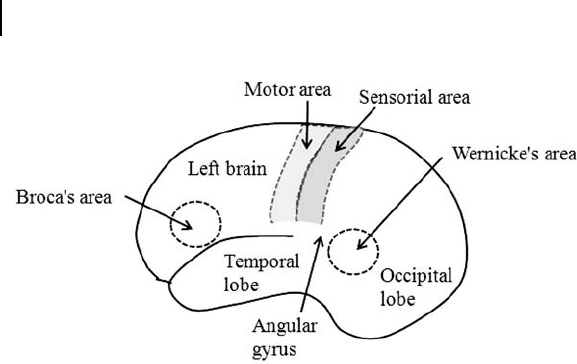
cognition. The occipital lobe is known to be involved in vision.
The temporal lobe in the left brain is related to speech, and, in
particular, Wernicke’s area, which is located there, is famous as the
part of the brain associated with the cognition of spoken and written
language.
The angular gyrus, located immediately posterior to Wernicke’s
area, is very much associated with information on meaning (Fig. 3.4).
According to experiments carried out by Wilder Graves Penfield
(1891–1976), the part of the brain involved in long-term memory
spreads over a wide area of the temporal lobe.
It is known that sexual arousal is generated when the temporal
lobe is stimulated, and the frontal lobe is active during both male
and female orgasms.
The cingulate gyrus of the frontal lobe is where researchers
believe self-consciousness resides. Self-consciousness is a function
by which an individual is capable of judging that an image in a mirror
is his/her own or someone else’s. Self-consciousness is also known
to be highly associated with Broca’s area, which is responsible
for speech functions. This suggests that being able to speak about
June 25, 2012 12:22 PSP Book - 9in x 6in 03-Junichi-Takeno-c03
26 Story of the Human Brain
Figure 3.4. Broca’s area and Wernicke’s area.
oneself could be an origin of self-consciousness. Broca’s area is
located in the frontal lobe. It has also been said that the area involved
in will exists in the prefrontal area of the frontal lobe.
The sensorial area and motor area exist in the region where the
parietal lobe and the occipital lobe meet. The sensorial area is where
the sub-areas corresponding to the tactile sensors of various parts
of the body exist. These sub-areas are cortices and receive signals
from respective tactile sensors. The cortices receiving signals from
the hands, fingers, lips, genitals, and toes occupy a relatively large
surface area (Fig. 3.4).
The sense of taste is associated with the cortex on the lower part
of the right frontal lobe.
The stimuli of smell directly enter the limbic system and activate
the lower part of the right frontal lobe.
The primary motor cortex lies at the center of the parietal lobe.
It sends signals to activate the motor functions of the body. The right
hemisphere’s primary motor cortex controls muscles on the left side
of the body, while that on the left controls muscles on the right side
of the body.
When a human wants to make a certain movement, the primary
motor cortex sends signals to contract muscles. At this point, there
may be information circulating between the vision and somatic
sensors in the body and the brain to monitor and make sure that
the desirable motion is achieved.
June 25, 2012 12:22 PSP Book - 9in x 6in 03-Junichi-Takeno-c03
Story of the Human Brain 27
The area involved in spatial cognition exists in the rear part of the
parietal lobe. Most of the occipital lobe is used for vision processing.
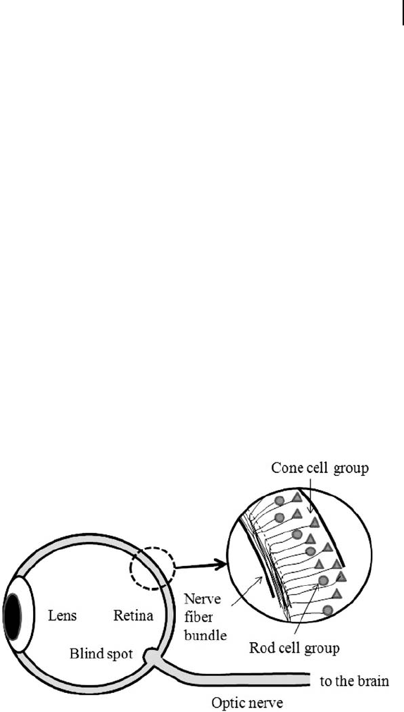
An image entering through the lenses of the eyes is inverted
when reaching the retinas. There are two types of photoreceptor
cells in the retina: cones and rods. The cone cells detect intensity
and the rod cells color information. The fovea in the retina is
where photoreceptors are most densely concentrated. Signals from
the photoreceptors, already image-processed on the retina to some
extent, leave the eyes and go to the brain via the part called the
blind spot and through a bundle of neural pathways. The blind spot
consists of a dense bundle of neural pathways and there are no
photoreceptors. The blind spot is therefore “invisible,” or cannot
respond to light stimulation. It is very interesting to note that the
photoreceptors are viewing light that has penetrated the layers of
nerve fiber bundles (from the back side of the nerve cables) since
the photoreceptors are located at the deepest part or bottom of the
retina (Fig. 3.5).
The neural pathways leave the right and left eyes and go to
the brain. On the way, they cross each other just once at a certain
location. This crossing point is called the optic chiasm.
Figure 3.5. Vision and nervous system.
June 25, 2012 12:22 PSP Book - 9in x 6in 03-Junichi-Takeno-c03
28 Story of the Human Brain
Because of this crossing, the neural pathways associated with the
right-side visual fields of both the right and left eyes go to the left
occipital lobe and the neural pathways associated with the left-side
visual fields of both eyes go to the right occipital lobe.
Optic chiasm has not been fully explained biologically. The author
supports the hypothesis that the optic chiasm has an advantage in
that even when if one hemisphere of the brain is damaged, the other
hemisphere of the brain keeps the functions of stereovision viable.
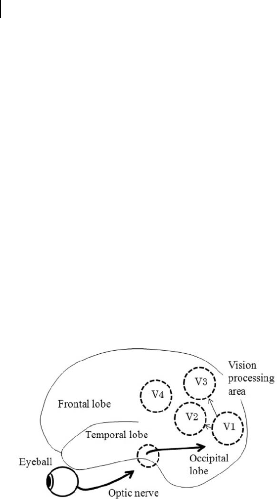
Visual information runs through the part of the brain called the
lateral geniculate body, which is a part of the thalamus and reaches
an area named the primary visual cortex (V1) in the right and left
occipital lobes.
V1 is like a projector that shows image information captured
by the eyes. Visual information is further transferred to other
visual areas called V2, V3, V4, and V5 for the brain to determine
the position of an object being observed. V2 processes three-
dimensional perception, V3 distance, and V5 motion information
(Fig. 3.6). The visual information passing through V1, V2, and V4
determines what the object is. V4 is responsible for processing color
information.
When the primary visual cortex of the occipital lobe has a
lesion, humans cannot see their outside world. It has been reported,
however, that even with the visual area damaged, part of the visual
Figure 3.6. Vision processing system.
..................Content has been hidden....................
You can't read the all page of ebook, please click here login for view all page.