
Sealing Behavior of Fracture in Cementitious Material with Micro-Focus X-ray CT
ABSTRACT. High strength and ultra low permeability concrete (HSULPC) has been studied for an alternative technology for long term confinement of Carbon14 (C-14) in geological disposal in geological disposal of transuranic radio waste (TRU waste). Thus knowledge of time-dependent fracturing and permeability is important. For cementitious materials, precipitation of calcium compound can occur on the surface. This can affect the time-dependent crack growth and permeability. In this study, the sealing behavior of cracks and pores in HSULPC submerged in water was observed by using micro-focus x-ray CT. It was shown that cracks and pores were sealed by precipitation of the calcium carbonate. Furthermore, it was shown that the sealed volume increased with increasing elapsed time and sealing rate was calculated. It is concluded that x-ray CT is useful for quantitative estimation of sealing of cracks and pores in cementitious material.
KEYWORDS: fracture sealing, cementitious material, micro-focus x-ray CT, image subtraction
1. Introduction
For geological disposal of radioactive wastes, the intensity of radioactivity of radionuclides can be reduced by engineered barriers such as bentonite buffer and by natural barriers such as a rock mass. If the repository of radioactive wastes is located in an area where the hydraulic gradient and the permeability are high, the retardation of migration of radionuclides by these barriers may not be enough. Therefore, the influences of nuclides with low adsorption, such as I-129 and C-14, will be large. In order to retard the migration, several alternative concepts of radioactive waste packages are being developed (Owada et al., 2005). It is planned that high-strength and ultra low-permeability concrete (HSULPC) will be used for a radioactive waste package for the geological disposal of transuranic radionuclides (TRU waste) (Kawasaki et al., 2005; Owada et al., 2005). Figure 1 shows a schematic illustration of this alternative concept using HSULPC. The aim of this scheme is to confine radionuclides, especially C-14, for a long term. It is supposed that C-14 is confined for 60,000 years, which corresponds to 10 times of the half-life of C-14. If a crack nucleates and propagates in HSULPC, however, the confining ability of the package can decline after crack growth for the long term even though the crack propagates slowly. Therefore, time-dependent fracturing of HSULPC has been studied (Nara et al., 2008).
In the case of cementitious materials, it is generally known that precipitation of calcium compounds on the surface occurs in water. Thus sealing of pores and cracks by precipitation is possible. This may affect the crack growth and permeability.
In this study, we investigated the sealing of cracks and pores. Specifically, the sealing of cracks and pores in HSULPC in water by calcium precipitation was observed using micro-focus x-ray CT. Then, the change of the sealed volume by precipitation with elapsed time was estimated.
2. Sample
In this study, HSULPC (made by Taiheiyo Consultant Corporation) was used as a sample. The composition of HSULPC is shown in Table 1. The bulk density was 2460 kg/m3. The uniaxial compressive strength was 203 MPa. The porosity was 5%. The water permeability coefficient was 4×10⁻¹⁹ m/s (Kawasaki et al., 2005).
In Figure 2, a specimen of HSULPC used in this study is shown. The cylindrical test pieces of HSULPC was split into two halves and set in the acrylic cylinder in the way that the width of two split fracture planes became 0.1 mm. The height of acrylic cylinder was 35 mm, and the diameter and height of split HSULPC specimen were approximately 13 mm and 15 mm, respectively.
Figure 1. Illustration of an alternative concept of radioactive waste package using HSULPC
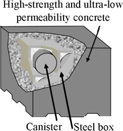
Figure 2. Split specimen of HSULPC
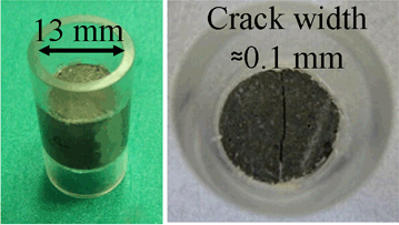
Table 1. The composition of HSULPC
| Contained amount [kg/m3] | |
| Low-heat Portland cement | 744∼1014 |
| Silica fume | 158∼496 |
| Fillers (fly ash, blast furnace slag, etc.) | 225∼541 |
| Aggregates | 631∼947 |
| Water-reducing admixture | 24 |
| Water | 180 |
3. Methodology
3.1. Observation method of precipitation
The micro-focus x-ray CT system made by Toshiba IT & Control Systems Corporation (TOSCANER 30900 µhd) was used to observe the sealing process in HSULPC specimens. The focal spot size of x-ray tube was 5 µm.
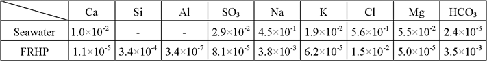
At first, the specimens were immersed in water and kept for a selected term at 20 degree Celsius. Specimens were stored in one of two types of water, i.e., simulated fresh reducing high pH type groundwater (FRHP) and simulated seawater. In Table 2, chemical composition of water is shown. Next, we took out a specimen from water at the desired term, dried it in a glove box filled with nitrogen gas and then observed it with x-ray CT. The observation was performed periodically over a 6 month term.
Table 2. Chemical composition of simulated seawater and simulated FRHP in mol/l
In x-ray CT observation, at first, the cone beam scanning was used for the 3D reconstruction of whole specimen with a resolution of 1024×1024 pixels in each slice. In this scanning mode, the slice thickness was set to be 0.04 mm and 402 slice images were obtained along the direction of the sample height. Then multi-slice scanning was conducted to obtain more precise images than those for cone beam scanning. This scanning mode can achieve a high resolution of 2048×2048 pixels which corresponds to a resolution of 7.6 µm. For the quantitative estimation by image analysis, images from multi-scanning were used.
3.2. Image analysis method
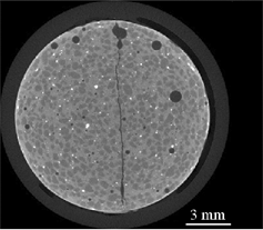
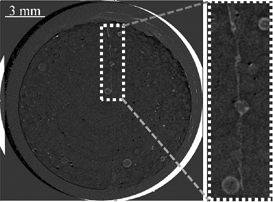
As mentioned before, images obtained from multi-slice scanning were used for image analysis because they had higher resolution. Specifically, the images of the samples obtained before the immersion were compared to those obtained after the immersion using the image subtraction method. In this study, the CT image before the immersion was defined as initial image. In Figure 3, an initial image is shown. It is necessary to correct the unavoidable alignment gap between each time of scanning so that the same regions of the initial and immersed core can be compared. For this purpose, affine transform was used. To obtain the optimal parameters for affine transform (displacements in two orthogonal directions (ux and uy) and rotation (ωxy)), following two steps were used. No distortion between initial image and images after immersion is assumed.
First, by comparing the initial image and post-immersion images for several representative points, approximate values of ux, uy and ωxy were obtained. Then, by changing these three parameters and calculating the cross correlation value, we found the maximum value of cross correlation and the optimal parameters of ux, uy and ωxy for affine transform. The value of cross correlation, Cc, is calculated as:
[1] ![]()
where f0 and fR are the CT values of individual pixels within the initial image and image after the immersion, respectively. Then the image subtraction analyses between the initial image and the images after the immersion were conducted.
Figure 3. An initial image of a specimen obtained by multi-slice scanning
4. Results
4.1. Results of observation
Images of x-ray CT of a specimen immersed in simulated FRHP obtained by cone beam scanning are shown in Figure 4 (radioactive waste management funding and research center (RWMC), 2009). In these images, dark colors correspond to cracks and pores.
Figure 4. X-ray CT images at the fracture obtained by cone beam scanning
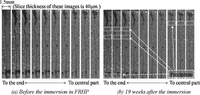
Figure 4(a) shows the initial images (before immersion) and Figure 4(b) shows images of the same positions of the specimen immersed in water for 19 weeks. It is clear that precipitation happened and the precipitate sealed the fracture plane. The same sealing behavior was found in the specimen immersed in simulated seawater. From observations obtained with x-ray CT, the sealed parts of the specimen were distinguishable in approximately 3 weeks of submergence in water. It was also found that sealing completed more quickly for the fracture with a smaller aperture. In Figure 4(b), sealing is apparent in the lower portion of the figure, yet it is not as complete as in the upper portion. Sealing was not observed in central part of the specimen. From these results, it is likely that the sealing rate decreased with the elapsed time after the immersion. The precipitate was mainly CaCO3 (RWMC, 2009) as determined by an Electron Prove Micro Analyser (EPMA).
4.2. Results of image analysis
In Figure 5, a result of image subtraction is shown. The parts with a lighter color indicate the sealed parts.
Figure 5. A result of image subtraction for a specimen immersed in simulated seawater
By using image subtraction analysis, the parts of the sample that were sealed by precipitation were revealed. As shown in Figure 5, the change of volume percentage of precipitate in a pore in case of simulated seawater was calculated by the result of image subtraction and threshold processing. Although the quantitative estimation for the pore resulted in success, there needs further study for the quantitative estimation of split part, because left hand side of Figure 5 has several pixels of non-zero grayscale values except in the area where precipitation of CaCO3 occurred. This is due to the inevitable difference of the scanning position. Since this difference was not considered in this study, it will be the future subject to conduct image analysis considering the difference of the scanning position.
5. Discussion
From the observation with x-ray CT, the sealed parts of fractures in HSULPC increased with elapsed time. It is important to estimate the sealed volume quantitatively in order to clarify the progress of sealing behavior. By using the result of image subtraction, we estimated the temporal change of the sealed volume. In particular, after using binary conversion to the subtraction image, we calculated the number of the pixel of sealed parts. Then, we determined the percentage of the sealed parts by using the following equation:
[2] ![]()
where Ps is the percentage of the sea the sealed parts, Ns is the number of the pixel Ps means the percentage of the sealed volume by precipitation. In Figure 6, the temporal change of Ps for one pore in the specimen immersed in simulated seawater is shown. From this figure, it is shown that Ps increased with elapsed time.
Figure 6. Temporal change of the sealed volume of a pore in specimen immersed in simulated seawater
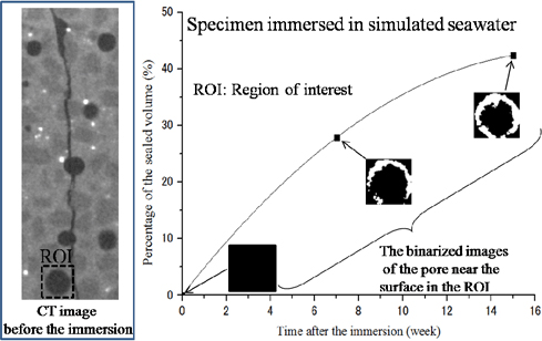
Thus Ps means the percentage of the sealed volume by precipitation. In Figure 6, the temporal change of Ps for one pore in the specimen immersed in simulated seawater is shown. From this figure, Ps increased with elapsed time. Additionally, it is shown that the change of Ps decreased. For precipitation of CaCO3, migration of water into and through HSULPC is necessary. Fracture and pore networks provide the principal pathways for fluid flow. If the pathway is partly sealed, water migration will be limited and precipitation may decrease. Therefore, it is considered that the decrease in the change of Ps (Figure 6) is caused by a temporal increase of the sealed volume by precipitation. Furthermore, the temporal increase of the sealed volume suggests that HSULPC possesses the property of self-sealing, which can delay fluid flow into and through these structures and ensure long-term integrity.
6. Conclusions
We have conducted the observation of sealing behavior in split HSULPC in water using x-ray CT and estimated the sealed volume with image analysis. The sealing behavior of cracks and pores by precipitation in HSULPC was visualized using a micro focus x-ray CT scanner. Sealing was observed mainly near the ends of the specimen. However, sealing was not observed in the central part of the specimen. The sealed volume increased with time while changes in sealed volume decreased with time due to a decrease of water migration in the specimen.
7. Acknowledgements
We would like to thank Radioactive Waste Management Funding and Research Center for the HSULPC samples used in the research project “Research and development of processing and disposal technique for TRU waste containing I-129 and C-14”, the Ministry of Economy, Trade and Industry and the Research Fellowships of the Japan Society for the Promotion of Science for Young Scientists.
8. References
Kawasaki, T., Asano, H., Owada, H. Otsuki, A., Yoshida, T., Matsuo, T., Shibuya, K., Takei, A., “Development of waste package for TRU-disposal (4) – Evaluation of confinement performance of TRU waste package made of High-Strength and Ultra Low-Permeability concrete”, Proc. GLOBAL 2005, No. 254, 2005.
Nara, Y., Mori, D., Owada, H. and Kaneko, K., “Study of subcritical crack growth and long-term strength for rock and cementitious material for radioactive waste disposal”, Proc. SHIRMS 2008, Vol. 2, p. 135-147, 2008.
Owada, H., Otsuki, A., Asano, H., “Development of waste package for TRU-disposal (1) – Concepts and performances”, Proc. GLOBAL 2005, No. 351, 2005.
Radioactive waste management funding and research center, Research and Development of Processing and Disposal Technique for TRU Waste Containing I-129 & C-14, Vol. 2, 2009.